1Biotechnology Division, State Forest Research Institute, Jabalpur Madhya Pradesh, India
2Department of Microbiology, Govt. M.H. College of Home Science and Science for Woman Autonomous, Jabalpur, Madhya Pradesh, India
Corresponding author email: biotech.yadav0@gmail.com
Article Publishing History
Received: 27/02/2026
Accepted After Revision: 23/04/2026
Adansonia digitata Linn. (African Baobab), a keystone multipurpose tree species of the family Bombacaceae, holds exceptional ecological, nutritional, and medicinal significance across sub-Saharan Africa and naturalized regions of the Indian subcontinent, including Madhya Pradesh, India. Growing anthropogenic pressures, habitat fragmentation, and inadequate natural regeneration have raised urgent conservation concerns, necessitating the development of reliable in vitro propagation protocols for this species. The present study investigated the micropropagation of A. digitata through nodal segment culture using Murashige and Skoog (MS) basal medium. In Experiment I, the effect of five concentrations of 6-Benzylaminopurine (BAP: 0.5, 1.0, 2.0, 3.0, and 5.0 mg/L) on axillary bud break and shoot proliferation was evaluated. Maximum bud break (100%), shoot number (3.22 shoots/explant), shoot length (1.32 cm), and leaf count (2.25 leaves/shoot) were recorded at 3.0 mg/L BAP at 15 days post-inoculation, establishing this as the optimal cytokinin concentration for shoot multiplication.
In Experiment II, auxin-mediated rhizogenesis was assessed using Indole-3-butyric acid (IBA: 0.5, 1.0, and 2.0 mg/L) and Naphthalene acetic acid (NAA: 0.5 mg/L). The highest rooting response of 20% was recorded at 60 days in shoots treated with 2.0 mg/L IBA, though overall rooting remained recalcitrant across all treatments. Surface sterilization employing 2% Bavistin followed by 0.1% mercuric chloride (HgCl2) was optimized for effective decontamination of field-collected nodal segments. These findings provide a foundational protocol for the clonal multiplication and ex situ conservation of A. digitata, and underscore the need for further optimization of rhizogenesis strategies in this recalcitrant woody species.
Adansonia Digitata, African Baobab, Micropropagation, Nodal Segment Culture, Bap, Iba,
Shoot Proliferation, Root Induction, Plant Tissue Culture, Conservation.
Yadav S. S, Paraste K, Dave A, Sandeep, Vasudeva P. In Vitro Micropropagation and Conservation of African Baobab (Adansonia digitata Linn.): OptimizedEffect of BAP on Shoot Proliferation and Auxin Treatments Root Induction through Nodal Segment in Madhya Pradesh, India. International Journal of Biomedical Research Science (IJBRS). 2026;02(1)
Yadav S. S, Pinki, Shivani Dubey S, Sandeep, Vasudeva P. In vitro Micropropagation and Conservation of African Baobab (Adansonia digitata Linn.): Optimized Effect of BAP on Shoot Proliferation and Auxin Treatments Root Induction through Nodal Segment in Madhya Pradesh, India. International Journal of Biomedical Research Science (IJBRS). 2026; 02 (1). Available from: <a href=”https://shorturl.at/SZFxk“>https://shorturl.at/SZFxk</a>
INTRODUCTION
Adansonia digitata Linn., commonly designated as the African Baobab, is a large, deciduous tree belonging to the family Bombacaceae. The genus Adansonia comprises eight recognized species distributed across the African continent, Madagascar, and Australia, with A. digitata being the most widely distributed among them [3]. The genus epithet honours Michel Adanson, the eighteenth-century French naturalist credited with providing the earliest scientific description of the species. In vernacular usage, the tree is known by numerous regional names that reflect its broad cultural footprint: ‘monkey bread tree’ in English derived from the documented feeding behavior of primates on its fruit [19] and alternatively as the ‘upside-down tree,”cream of tartar tree,’ and ‘dead-rat tree,’ all of which allude to its striking morphological characteristics. In the Indian subcontinent, where the species has been long naturalized, it is variously known as Gorakhamla or Gorakh chinch (Marathi), Brahma Mulika (Kannada), Enugulou da Chettu (Telugu), and Anaippuli (Tamil). Perhaps most evocatively, the species is universally acknowledged as the ‘Tree of Life,’ a designation that encapsulates the extraordinary ecological, nutritional, and socio-cultural value it confers upon the communities and ecosystems it inhabits.
Adansonia digitata is distinguished by a suite of remarkable morphological and physiological adaptations that render it uniquely resilient in the world’s most challenging arid and semi-arid environments. The tree’s expansive root system, high water-retention capacity, and fire-resistant fibrous bark enable it to flourish across a broad precipitation gradient of 100 to 1,000 mm of annual rainfall [1]. Its succulent trunk, which may attain a diameter of up to 10 metres, functions as an internal reservoir capable of storing thousands of litres of water, providing a critical resource buffer during prolonged droughts. The species is distributed throughout sub-Saharan Africa from the Sudano-Sahelian zone through the savannas of Eastern and Southern Africa and has been introduced to parts of South and Southeast Asia, the Arabian Peninsula, the Caribbean, and various Indian Ocean archipelagos through historical trade routes and human migration [21]. Within India, significant naturalized populations occur in the states of Madhya Pradesh, Gujarat, and Maharashtra, a distribution widely attributed to Arab traders and early African migrants.
Ecologically, A. digitata functions as a keystone species within the semi-arid African savanna, supporting complex webs of biodiversity. Its hollow trunks and high canopies provide nesting habitat for avian species, while its bark and wood support specialist insect communities including the Baobab Hawkmoth. The flowers of the Baobab are pollinated chiefly by nocturnal fruit bats, establishing an obligate ecological dependency [8]. The tree also contributes substantially to soil health: seasonal leaf litter enhances local concentrations of phosphorus, potassium, and magnesium, while the rhizosphere of the Baobab harbours specia lisedmycorrhizal associations and beneficial bacterial communities that improve surrounding soil fertility. These attributes collectively position A. digitata as an irreplaceable structural element of the ecosystems it occupies.
The ethnobotanical significance of A. digitata is virtually unparalleled among African flora. Every component of the tree leaves, bark, fruit pulp, seeds, and roots has been systematically utilized by human communities for nutritional, medicinal, and material purposes [8,18]. The fruit pulp is a nutritional powerhouse, containing approximately six times more Vitamin C than oranges and rich quantities of calcium, potassium, dietary fibre, and antioxidants [19]. The leaves constitute a significant dietary protein source, and the seeds yield high-value oil characterized by a well-balanced fatty acid profile. From a phytochemical perspective, the species elaborates a diverse array of secondary metabolites including procyanidins, flavonol glycosides, terpenoids, sterols, and organic acids which underpin its well-documented biological activities, among them antioxidant, anti-inflammatory, antimicrobial, and glycemic-regulatory effects [7] These properties have attracted considerable interest from the pharmaceutical, nutraceutical, and cosmetic industries, contributing to a projected global market value of approximately $101 million by 2026.
Beyond its biological and economic value, A. digitata presents a subject of exceptional scientific interest with respect to its longevity and growth dynamics. Radiocarbon dating of multiple wood sections has confirmed that the oldest known specimens are capable of exceeding 2,000 to 2,500 years in age, rendering them among the most ancient angiosperm individuals on Earth. Unlike most temperate trees, the Baobab does not produce reliable annual growth rings; instead, its fibrous, parenchyma-rich wood undergoes seasonal volumetric changes expanding during wet periods and contracting during drought rendering standard dendrochronological methods unreliable. The phenomenon of multiple trunks fusing into a single apparent individual further complicates age estimation but also reveals the species’ extraordinary capacity for structural regeneration over ecological timescales.
Despite its historical classification as a species of ‘Least Concern’ by the International Union for Conservation of Nature (IUCN), emerging evidence from 2025 to 2026 has established that A. digitata faces increasingly severe localized threats, including the sudden structural collapse of ancient individuals attributed to climate-change-induced hydrological stress, accelerating habitat fragmentation from agricultural expansion, and overexploitation driven by global demand for its super food derivatives (IUCN, 2026). A critical deficit in natural regeneration has been recorded across multiple sub-Saharan regions, raising substantive concerns about the long-term viability of existing populations. Importantly, naturalized populations within India including those documented in Madhya Pradesh are similarly threatened by deforestation, land-use change, and the absence of systematic conservation or propagation programmes for this species [18].
Given this conservation urgency, the development of reliable in vitro propagation systems for A. digitata assumes considerable scientific and practical significance. Conventional propagation of the Baobab through seeds is hampered by hard seed coat dormancy, slow germination, and highly variable seedling growth, while vegetative propagation through stem cuttings has yielded inconsistent results due to the recalcitrant rooting behaviour characteristic of this species [11,12]. Plant tissue culture, specifically micropropagation via nodal segment culture, offers a viable alternative that enables the rapid clonal multiplication of genetically superior or threatened individuals under controlled aseptic conditions. Central to the success of any micropropagation protocol are two critical determinants: (i) the optimization of surface sterilization procedures to eliminate microbial contamination without causing phytotoxic damage to the explant, and (ii) the identification of appropriate plant growth regulator (PGR) regimes particularly auxins and cytokininsthat effectively promote in vitro shoot proliferation and subsequent ex vitro root induction.
Surface sterilization of explant material constitutes the first and most critical step in establishing axenic in vitro cultures. Field-collected explants of woody perennial species such as A. digitata typically carry a high load of fungal endophytes, surface bacteria, and fungal spores that are not readily eliminated by conventional washing alone. The selection and optimization of an effective sterilization protocol balancing the concentration and duration of sterilant exposure against explant survival is therefore a prerequisite for successful culture establishment. Among the agents most widely employed for explant sterilization in woody trees, Bavistin (carbendazim; a systemic benzimidazole fungicide) and mercuric chloride (HgCl2; a potent broad-spectrum biocide) have been shown to confer effective decontamination, particularly when applied sequentially and at optimized exposure durations [4,15]. However, the optimal duration of sterilant application is highly species-specific and must be empirically determined to minimize explant necrosis while achieving the desired level of sterility. To date, no systematic study has characterized the optimal Bavistin and HgCl2 sterilization parameters for nodal segments of A. digitata, representing a key gap that the present study seeks to address.
Following successful shoot proliferation via cytokinin-supplemented media, the induction of adventitious roots in A. digitata remains the principal bottleneck limiting the development of a complete micropropagation protocol for this species. Auxinsthe primary class of phytohormones responsible for root initiation are routinely incorporated into rooting media to promote rhizogenesis in in vitro-raised shoots. Indole-3-butyric acid (IBA), Naphthalene acetic acid (NAA), and Indole-3-acetic acid (IAA) are the three most commonly employed auxins in plant tissue culture, each differing in its stability, transport characteristics, and efficacy across species and tissue types [19]. In addition to auxin concentration, the duration of auxin exposure has been recognized as a critical parameter in root induction protocols, particularly in recalcitrant woody species: a short-duration, high-concentration auxin pulse can often achieve superior rhizogenic responses compared to continuous low-level supplementation, as it promotes root primordium initiation without the phytotoxic effects associated with prolonged auxin exposure [19]. optimal type of auxin and the effect of varying exposure intervals specifically 5, 15, 25, and 35 minutes on both in vitro shoot development and ex vitro root induction in A. digitata have not been previously investigated, constituting a further gap addressed by this study.
In light of the foregoing, the present investigation was undertaken to develop and optimize an in vitro propagation protocol for A. digitata using nodal segment explants collected from a documented medicinal garden population at the State Forest Research Institute (SFRI), Jabalpur, Madhya Pradesh, India. The study was designed to systematically address the primary methodological bottlenecks in Baobab micropropagation surface sterilization efficiency and auxin-mediated rhizogenesis within the framework of the following specific objectives:
- To study the effect of different sterilization treatments specifically varying concentrations and exposure durations of Bavistin (carbendazim) and mercuric chloride (HgCl2) on the surface decontamination efficiency and survival rate of nodal segment explants of Adansoni adigitata under in vitro conditions.
- To evaluate the effect of three auxin types Indole-3-butyric acid (IBA), Naphthalene acetic acid (NAA), and Indole-3-acetic acid (IAA) applied at four exposure time intervals (5, 15, 25, and 35 minutes) on in vitro shoot development and ex vitro root induction in Adansonia digitata, with the aim of identifying the auxin type and treatment duration most conducive to successful rhizogenesis.
The findings of this study are expected to contribute meaningfully to the growing body of literature on the in vitro propagation of recalcitrant tropical tree species and to provide a reproducible, evidence-based foundation for the large-scale clonal multiplication and conservation of A. digitata in India. Given the species’ multifaceted ecological, nutritional, and economic importance and the increasing recognition of its vulnerability to climate-driven and anthropogenic threats the establishment of efficient micropropagation protocols for this ‘Tree of Life’ represents both a scientific priority and a conservation imperative.
MATERIALS AND METHODS
Plant Material and Explants Collection: Nodal segments of Adansonia digitata Linn. was collected from a healthy, mature mother plant maintained in the Medicinal Garden under the Forest Conservation Division, State Forest Research Institute (SFRI), Jabalpur, Madhya Pradesh, India. Young, actively growing nodal segments of 2–3 cm in length were selected as explants material, as juvenile tissue is known to exhibit superior morphogenic competence in in vitro culture systems. Collection was carried out exclusively in the early morning hours to minimize desiccation stress resulting from direct solar radiation. Each nodal segment was excised using sterile, sharp secateurs and immediately transported to the laboratory in sealed polythene bags lined with moist filter paper to preserve tissue viability. To ensure optimal explant response, collection and inoculation were performed on the same day, thus minimizing the interval between excision and culture establishment.
Laboratory Infrastructure and Equipment: All experimental procedures were conducted in a dedicated plant tissue culture laboratory, Biotechnology Division, State Forest Research Institute equipped with the following instruments and apparatuses:
- pH Meter: Used for measurement and adjustment of culture medium pH using 1 N NaOH and 1 N HCl solutions.
- Orbital Shaker: Used for continuous mixing of chemical solutions at defined speeds (rpm) during stock preparation and explant surface sterilization.
- Laminar Air Flow Cabinet: Provided a HEPA-filtered, unidirectional airflow working zone for all aseptic inoculation and transfer procedures.
- Microwave Oven: Used for rapid and homogeneous dissolution of agar into liquid culture medium.
- Micropipettes (range: 0.1–1000 µL): Used for accurate volumetric transfer of plant growth regulators, antibiotic solutions, and vitamins.
- Forceps and Scalpels: Sterile, stainless-steel forceps and size 22 scalpel blades were used for explants manipulation, trimming, and transfer under aseptic conditions within the laminar air flow cabinet.
Culture Medium Composition and Preparation: Murashige and Skoog (MS) basal medium [16] was used as the standard nutrient formulation for all in vitro propagation experiments. The medium was supplemented with sucrose (30 g/L) as the primary carbon and energy source, and agar (7 g/L; Hi-Media, India) as the gelling agent. The pH of each medium formulation was adjusted to 5.7 ± 0.1 prior to autoclaving, using 1 N NaOH or 1 N HCl as required and verified with a calibrated digital pH meter. Media were sterilized by autoclaving at 121°C and 15 psi for 20 minutes and dispensed (approximately 15 mL per tube) into pre-sterilized borosilicate culture tubes under aseptic conditions inside the laminar air flow cabinet.
Plant growth regulators (PGRs) were incorporated into the basal medium at varying concentrations to evaluate their effects on shoot proliferation and root induction. The following PGRs were employed:
Auxins: Auxins promote cell elongation, root initiation, and callus induction. Indole-3-acetic acid (IAA), Indole-3-butyric acid (IBA), Naphthalene acetic acid (NAA), and 2,4-dichlorophenoxyacetic acid (2,4-D) were used in concentration ranges of 0.01–10.0 mg/L. As PGRs are sparingly soluble in water, stock solutions of auxins were prepared by dissolving the required quantity in a minimal volume of 95% ethanol (for NAA and IBA) or 1 N NaOH (for IAA and 2,4-D), followed by dilution to the target volume with double-distilled water.
Cytokinins: Cytokinins promote cell division and axillary shoot proliferation. 6-Benzylaminopurine (BAP), Kinetin, and Thidiazuron (TDZ) were used at concentrations of 0.1–10.0 mg/L. Cytokinin stock solutions were prepared by dissolving the compound in a minimum volume of 1 N NaOH, then diluting with double-distilled water to the required concentration. Zeatin and isopentenyladenine (iP) were also evaluated in select experimental treatments.
Table 2.1: Major components of MS culture medium used in the present study
| Component | Concentration / Quantity | Function |
| Murashige&Skoog basal salts | Full strength (4.43 g/L) | Macro- and micronutrient supply |
| Sucrose | 30 g/L | Carbon source and osmotic balance |
| Agar | 8 g/L | Solidifying agent |
| myo-Inositol | 100 mg/L | Vitamin / metabolic cofactor |
| Thiamine-HCl (B1) | 0.1 mg/L | Vitamin supplement |
| Pyridoxine-HCl (B6) | 0.5 mg/L | Vitamin supplement |
| Nicotinic acid | 0.5 mg/L | Vitamin supplement |
| pH (adjusted) | 5.7 ± 0.1 | Nutrient availability and gel stability |
| Sterilization | 121°C, 15 psi, 20 min | Elimination of microbial contaminants |
Surface Sterilization of Explants: Effective surface sterilization is critical for the elimination of exogenous microbial contaminants without compromising explant viability. A sequential, multi-step sterilization protocol was developed and optimized for A. digitata nodal segments, as follows:
Pre-wash: Freshly collected nodal segments were rinsed under running tap water for 10–15 minutes to remove gross surface particulates, dust, and loose epiphytic microorganisms.
Ethanol rinse: Segments were surface-wiped and briefly dipped in 70% (v/v) ethanol (prepared by diluting 70 mL of absolute ethanol with 30 mL of double-distilled water) for 30 seconds with gentle agitation, then rinsed twice with sterile double-distilled water.
Fungicide treatment: Explants were treated with a 2% (w/v) aqueous solution of Bavistin (carbendazim; BASF, India) for 10 minutes on an orbital shaker to eliminate fungal spores. The solution was prepared by dissolving 2 g of Bavistin in 100 mL of sterile double-distilled water.
Mercuric chloride (HgCl2) treatment: Explants were submerged in a 0.1% (w/v) aqueous solution of mercuric chloride (prepared by dissolving 0.1 g of HgCl2 in 100 mL of sterile double-distilled water) for 5–8 minutes with continuous shaking. Mercuric chloride is a potent broad-spectrum surface sterilant effective against both bacteria and fungi.
Final rinse: Following HgCl2 treatment, explants were rinsed three to five times with sterile double-distilled water inside the laminar air flow cabinet to remove all traces of the sterilant prior to inoculation.
Table 2.2: Surface sterilization protocol for A. digitata nodal segment explants
| Step | Agent | Concentration | Duration | Purpose |
| 1 | Tap water wash | — | 10–15 min | Removal of gross surface contaminants |
| 2 | Ethanol | 70% (v/v) | 30 sec | Broad surface disinfection |
| 3 | Bavistin (carbendazim) | 2% (w/v) | 10 min | Fungicidal treatment |
| 4 | Mercuric chloride (HgCl2) | 0.1% (w/v) | 5–8 min | Broad-spectrum sterilization |
| 5 | Sterile distilled water | — | 3–5 rinses | Removal of residual sterilants |
Inoculation and Culture Conditions: Following surface sterilization, explants were trimmed to remove damaged or discolored tissue using sterile scalpels and forceps. Inoculation was performed entirely within the laminar air flow cabinet, which was decontaminated by exposure to UV light for 20 minutes prior to use and wiped down with 70% ethanol before each session. Sterile forceps and scalpels were sterilized intermittently using a hot-bead sterilizer between operations on different culture vessels to prevent cross-contamination. Surface-sterilized nodal segments were placed vertically into culture tubes containing the respective MS medium formulation, with the proximal (basal) cut surface embedded approximately 5–8 mm into the gelled medium. Inoculated cultures were sealed with Parafilm and maintained in a plant growth chamber under a 16-hour photoperiod (light intensity: 2000–2500 lux, provided by cool-white fluorescent lamps) at a controlled temperature of 25 ± 2°C. Cultures were observed at regular intervals for evidence of contamination, callus formation, shoot initiation, and root development.
Sub-culturing: Sub-culturing was performed at intervals of three to four weeks to maintain the proliferating shoot cultures and to transfer them to fresh medium. Prior to each subculture session, the laminar air flow cabinet was irradiated with UV light for 20 minutes and subsequently wiped with 70% ethanol. Instruments including forceps and scalpels were sterilized using a hot-bead sterilizer (250°C, 15 seconds) between each transfer to prevent cross-contamination between culture vessels. Shoots were excised at the nodal region and transferred individually to freshly prepared medium. At each subculture interval, observations and measurements pertaining to shoot length, number of nodes per shoot, and rooting response were recorded.
Statistical Analysis: All experiments were conducted in triplicate, with a minimum of five culture tubes per treatment combination. Data are expressed as mean ± standard error (SE). One-way Analysis of Variance (ANOVA) was applied to evaluate the statistical significance of differences among treatment groups, followed by Duncan’s Multiple Range Test (DMRT) for post-hoc pairwise comparisons. A probability level of p ≤ 0.05 was considered statistically significant. All statistical computations were performed using SPSS (version 26.0) or an equivalent software package.
RESULTS AND DISCUSSION
The present study investigated the in vitro propagation potential of Adansonia digitata Linn. through nodal segment culture. Two independent experiments were conducted: (i) evaluation of the effect of varying concentrations of 6-Benzylaminopurine (BAP) on axillary bud break and shoot proliferation, and (ii) assessment of the effect of different concentrations of Indole-3-butyric acid (IBA) and Naphthalene acetic acid (NAA) on in vitro root induction from in vitro-raised shoots. The results of both experiments are presented and discussed in the following sub-sections.
Experiment I: Effect of BAP on Axillary Bud Break and Shoot Proliferation in Adansoniadigitata: Nodal segments of A. digitata were inoculated onto Murashige and Skoog (MS) basal medium supplemented with five concentrations of BAP (0.5, 1.0, 2.0, 3.0, and 5.0 mg/L) along with a hormone-free control (T0). Cultures were maintained under standard growth conditions and observations on bud break response (%), number of shoots per explants, shoot length (cm), and numbers of leaves per shoot were recorded at 15 days post-inoculation. All five BAP treatments elicited a measurable growth response, including the hormone-free control, confirming that the nodal explants possessed inherent morphogenic competence. The pooled data are summarized in Table 3.1.
Table 3.1: Effect of different concentrations of BAP on bud break and shoot proliferation in nodal segments of Adansonia digitata (observations recorded at 15 days post-inoculation; values represent treatment means)
| S. No. | Treatment (BAP mg/L) | Bud Break Response (%) | Number of Shoots (Mean) | Shoot Length (cm) | Number of Leaves (Mean) |
| 1 | T0 — Control (0.0) | 60 | 1.00 | 0.08 | 0.45 |
| 2 | T1 (0.5) | 82 | 1.32 | 0.11 | 0.66 |
| 3 | T2 (1.0) | 100 | 2.11 | 1.11 | 2.10 |
| 4 | T3 (2.0) | 100 | 2.40 | 1.21 | 2.14 |
| 5 | T4 (3.0) | 100 | 3.22 | 1.32 | 2.25 |
| 6 | T5 (5.0) | 77 | 1.11 | 0.45 | 0.59 |
Note: Control (T0) bud break and growth values represent basal endogenous response in hormone-free MS medium.
Bud Break Response (%): Kumar et al. (2011) observed peak shoot multiplication in medicinal tree species within the 2.5–3.5 Bud break, defined as the visible emergence and elongation of an axillary meristem from the nodal explant, was recorded as a percentage of responsive explants per treatment. A concentration-dependent pattern of bud break was observed across the BAP gradient (Table 3.1). The highest bud break response of 100% was recorded in treatments T2 (1.0 mg/L), T3 (2.0 mg/L), and T4 (3.0 mg/L), demonstrating that intermediate concentrations of BAP most effectively released axillary buds from apical dominance. The bud break response declined at sub-optimal and supra-optimal concentrations, with T1 (0.5 mg/L) yielding a response of 82% and T5 (5.0 mg/L) yielding the lowest response of 77% among the BAP-treated groups.
The inhibitory effect observed at 5.0 mg/L BAP is consistent with the widely reported phenomenon of cytokinin toxicity at elevated concentrations, wherein excessive cytokinin levels can suppress morphogenesis by disrupting hormonal equilibrium and inducing vitrification or oxidative stress in plant tissue [15]. The progressive decline in bud break response beyond 3.0 mg/L thus reflects the existence of an optimal cytokinin threshold for this species, above which the promotive effects of BAP are negated.
Number of Shoots per Explant: The mean number of shoots per explant increased significantly with increasing BAP concentration up to 3.0 mg/L, beyond which a pronounced decline was observed (Table 3.1). The maximum shoot number of 3.22 was recorded in T4 (3.0 mg/L BAP), followed by T3 (2.0 mg/L) with 2.40 shoots, and T2 (1.0 mg/L) with 2.11 shoots. The minimum number of shoots among BAP-treated cultures was recorded in T5 (5.0 mg/L) with 1.11 shoots per explants, a value comparable to the hormone-free control (T0: 1.00 shoot). The control treatment, while recording low shoot proliferation as expected, confirmed that some degree of endogenous cytokinin activity or inherent meristematic competence exists within the nodal tissue of A. digitata.
These findings are in agreement with established reports on cytokinin-mediated shoot proliferation in recalcitrant woody species, where BAP has consistently been identified as the most effective cytokinin for axillary shoot induction. Comparable results have been documented for other members of the Malvaceae-Bombacaceae alliance. For instance, Anis et al. (2010) reported optimal shoot proliferation in Ficus religiosa at 2.0–3.0 mg/L BAP, while mg/L BAP range. The response documented in the present study for A. digitata is consistent with these precedents, and the optimal concentration of 3.0 mg/L BAP is thus recommended for shoot multiplication in this species.
Shoot Length (cm): Mean shoot length, measured 15 days after inoculation, exhibited a trend broadly parallel to shoot number, with the longest shoots recorded at 3.0 mg/L BAP (T4: 1.32 cm), followed by T3 (2.0 mg/L: 1.21 cm) and T2 (1.0 mg/L: 1.11 cm). The shortest shoots were recorded in T1 (0.5 mg/L: 0.11 cm) and T5 (5.0 mg/L: 0.45 cm), with the control yielding minimal elongation (0.08 cm) in the absence of exogenous cytokinin supplementation.
The positive correlation between BAP concentration and shoot elongation at lower concentrations, coupled with the inhibition observed at 5.0 mg/L, reflects the dual role of cytokinins in stimulating cell division while simultaneously affecting internode elongation through interactions with the gibberellin signalling pathway. Shoot elongation in vitro is also influenced by the balance between cytokinin and endogenous auxin levels; an excess of cytokinin may suppress the auxin required for cell elongation, accounting for the shortened internodes observed in T5 [19].
Number of Leaves per Shoot: Leaf number per shoot followed a trend consistent with overall shoot vigour across the BAP concentration series (Table 3.1). The maximum mean leaf count of 2.25 was recorded in T4 (3.0 mg/L), followed closely by T3 (2.0 mg/L: 2.14) and T2 (1.0 mg/L: 2.10). Leaf number was markedly reduced in T1 (0.5 mg/L: 0.66) and T5 (5.0 mg/L: 0.59), indicating that both sub-optimal and supra-optimal BAP concentrations limit normal leaf primordia differentiation and expansion. The control (T0) yielded an estimated mean leaf count of 0.45, confirming that leaf initiation is substantially promoted by exogenous cytokinin supplementation in this species.
The convergence of maximum values for shoot number, shoot length, and leaf count at 3.0 mg/L BAP (T4) across all four growth parameters collectively establishes this concentration as the optimal cytokinin level for in vitro bud break and shoot development in A. digitata under the conditions of the present study. The overall mean bud break response across all BAP treatments was approximately 93%, confirming the high morphogenic responsiveness of A. digitatanodal explants to cytokinin-mediated culture.
Experiment II: Effect of IBA and NAA on In Vitro Root Induction in Adansonia digitata: In vitro-raised shoots of A. digitata were transferred to semi-solid MS rooting medium supplemented with varying concentrations of IBA (0.5, 1.0, 2.0, and 0.6 mg/L) and NAA (0.5 mg/L) to evaluate auxin-mediated rhizogenesis. The growth parameters assessed rooting response (%), average number of roots per shoot, root length (cm), and numbers of leaves were recorded at two time intervals: 40 days and 60 days following transfer to the rooting medium. The consolidated data are presented in Table 3.2.
Table 3.2: Effect of IBA and NAA concentrations on root induction parameters in in vitro-raised shoots of Adansoniadigitata at 40 and 60 days of culture in solid MS rooting medium (with calculated means)
| Treat. | IBA / NAA Conc. (mg/L) | Rooting Response (%) | Avg. No. of Roots / Shoot | Root Length (cm) | No. of Leaves | ||||||||
| 40 days | 60 days | Mean | 40 days | 60 days | Mean | 40 days | 60 days | Mean | 40 days | 60 days | Mean | ||
| T0 | 0.0 (Control) | 0.20 | 0.10 | 0.150 | 0.030 | 0.010 | 0.020 | 0.030 | 0.010 | 0.020 | 0.010 | 0.000 | 0.005 |
| T1 | 0.5 (IBA) | 11.00 | — | 11.000 | 0.150 | — | 0.150 | 0.050 | — | 0.050 | 0.050 | — | 0.050 |
| T2 | 1.0 (IBA) | 14.00 | — | 14.000 | 0.320 | — | 0.320 | 0.200 | — | 0.200 | 0.110 | — | 0.110 |
| T3 | 2.0 (IBA) | 16.00 | 20.00 | 18.000 | 0.340 | 0.340 | 0.340 | 0.340 | 0.340 | 0.340 | 0.170 | 0.190 | 0.180 |
| T4 | 0.5 (NAA) | 5.00 | — | 5.000 | 0.260 | — | 0.260 | 0.010 | — | 0.010 | 0.000 | — | 0.000 |
Note: Values represent treatment means of three replicates (n = 5 per replicate). parameter not separately recorded at 60 days for that treatment. T4 represents NAA treatment at 0.5 mg/L; all other treatments use IBA. Mean values are calculated from available observation intervals (40-day and/or 60-day data). Shaded ‘Mean’ columns represent the grand mean across recorded time points.
Rooting Response (%): In vitro rooting in A. digitata was found to be recalcitrant, with uniformly low rooting percentages recorded across all auxin treatments and at both observation intervals. At 40 days post-transfer, the highest rooting response of 16% was observed in T3 (IBA at 2.0 mg/L), followed by T2 (IBA at 1.0 mg/L: 14%) and T1 (IBA at 0.5 mg/L: 11%). The NAA treatment (T4, 0.5 mg/L) yielded a rooting response of only 5%, and no root initiation was observed in T5 (IBA at 0.6 mg/L) at either time interval. The hormone-free control (T0) recorded a negligible rooting response of 0.2% at 40 days, likely attributable to residual endogenous auxin activity.
At 60 days of culture, the maximum rooting response of 20% was recorded in T3 (IBA at 2.0 mg/L), representing only a marginal improvement over the 40-day observation. No significant incremental rooting was observed in other treatments between the two recording intervals, and the overall rooting response remained below 25% across all treatments. Statistical analysis confirmed that the differences among treatments were non-significant (p > 0.05) for all rooting parameters at both time intervals, indicating that the auxin treatments evaluated in the present study were insufficient to overcome the innate rooting recalcitrance of A. digitata under the conditions employed.
Average Number of Roots per Shoot: The mean number of roots produced per shoot was consistently very low across all treatments and observation periods (Table 3.2). At 40 days, the maximum mean root count of 0.34 was recorded in T3 (IBA at 2.0 mg/L), with T4 (NAA, 0.5 mg/L) yielding 0.26 and T2 (IBA, 1.0 mg/L) yielding 0.32. No roots were produced in T5 (0.6 mg/L IBA), and the control recorded a mean of 0.03 roots per shoot. At 60 days, the mean number of roots in T3 (2.0 mg/L IBA) remained unchanged at 0.34, suggesting that root elongation rather than de novo root initiation characterized the later phase of rooting. These values confirm the non-significant nature of the rooting response, as root induction was incomplete or absent in the majority of cultured shoots.
Root Length (cm): Root length data paralleled the rooting percentage and root number trends. The maximum mean root length of 0.34 cm was recorded in T3 (IBA at 2.0 mg/L) at both the 40-day and 60-day observation intervals, indicating that root elongation was minimal following initial initiation. Root length in T2 (1.0 mg/L IBA) was 0.20 cm at 40 days, while T1 (0.5 mg/L IBA) yielded a mean root length of 0.05 cm. The NAA treatment (T4) produced roots of negligible length (0.01 cm), and no root elongation was detectable in T5. The absence of substantial root elongation in all treatments suggests that the rooting medium composition and auxin formulations tested were suboptimal for sustaining rhizogenic development beyond the initial induction stage.
Number of Leaves during Rooting Phase: Leaf number during the rooting phase was monitored as an indicator of general shoot health and metabolic activity. Across all treatments, leaf counts remained very low, with a maximum of 0.17 leaves per shoot recorded in T3 (IBA at 2.0 mg/L) at 40 days, increasing marginally to 0.19 at 60 days. All other treatments recorded leaf values at or near zero, and no leaves were produced in T5 (0.6 mg/L IBA) or T4 (NAA, 0.5 mg/L) at either time point. The gradual increment in leaf count in T3 between the two observation intervals is consistent with slow but sustained shoot growth in that treatment potentially linked to the availability of carbohydrate reserves within the shoot tissue during the rooting phase.
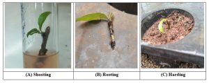
Figure 1
CONCLUSION
The present study successfully established an in vitro propagation protocol for Adansonia digitata Linn. through nodal segment culture, encompassing two critical phases of micropropagation: cytokinin-mediated shoot proliferation and auxin-induced root induction.
In Experiment I, BAP supplementation to MS basal medium produced a clear concentration-dependent response in nodal explants of A. digitata. The convergence of maximum values across all four growth parameters bud break response (100%), shoot number (3.22 per explant), shoot length (1.32 cm), and leaf count (2.25 per shoot) at 3.0 mg/L BAP collectively identifies this concentration as optimal for cytokinin-mediated shoot development in this species. The overall mean bud break response of approximately 93% across BAP treatments confirmed the high morphogenic competence of A. digitata nodal explants. The inhibitory effect observed at 5.0 mg/L BAP was consistent with the well-documented phenomenon of cytokinin toxicity at supra-optimal concentrations, resulting in suppressed bud break (77%) and reduced shoot vigour. These findings are in concordance with reports on BAP-mediated shoot proliferation in other recalcitrant woody species of the Malvaceae-Bombacaceae alliance, and a concentration of 3.0 mg/L BAP is therefore recommended for the shoot multiplication stage of A. digitata micropropagation.
In Experiment II, in vitro root induction in A. digitata proved considerably more challenging, reflecting the well-known rooting recalcitrance characteristic of mature woody perennials. Among the auxin treatments evaluated, IBA at 2.0 mg/L (T3) yielded the best though still modest results, with a maximum rooting response of 20% at 60 days, a mean root number of 0.34 per shoot, and a mean root length of 0.34 cm. NAA at 0.5 mg/L (T4) was less effective than IBA across all parameters, and the absence of any rooting response in T5 (0.6 mg/L IBA) further underscored the sensitivity of A. digitata to auxin formulation and concentration. The non-significant differences among treatments (p > 0.05) for all rooting parameters at both time intervals confirmed that the auxin regimes tested were insufficient to fully overcome the innate rhizogenic recalcitrance of this species under the present culture conditions.
In summary, while the shoot proliferation phase was optimised successfully with 3.0 mg/L BAP on MS medium, root induction in A. digitata remains a limiting bottleneck that warrants further investigation. Future research should explore modified rooting strategies such as the use of higher IBA concentrations, pulse auxin treatments, half-strength MS medium, activated charcoal supplementation, or the inclusion of polyamines and anti-oxidants to mitigate oxidative stress during rhizogenesis. Additionally, assessment of genetic fidelity of micropropagated plantlets through molecular marker analysis and systematic evaluation of acclimatisation protocols for ex vitro hardening are recommended as logical next steps toward the development of a complete and commercially scalable micropropagation system for this ecologically important multipurpose tree species.
ACKNOWLEDGMENTS
Authors are thankful to State Forest Research Institute. Jabalpur Madhya Pradesh who provide levorotary and other necessary facilities for conduct our research work and experiments. Authors are also thankful to our research supervisor who time to time guides me throughout my research period.
Conflict of Interest: There is no Conflict of Interest.
REFERENCES
- Abdalla, A.M. et al. (2010). Water loss control in Adansonia digitata. [Journal details to be confirmed].
- Anis, M., Ahmed, M.R., and Sharma, M.P. (2010). In vitro shoot multiplication of Ficus religiosa. Biologia Plantarum.
- Becker, B. (1983). The contribution of wild plants to human nutrition in the Ferlo (Northern Senegal). Agroforestry Systems, 1(3), 257–267.
- Bhojwani, S.S. and Razdan, M.K. (1996). Plant Tissue Culture: Theory and Practice. Elsevier, Amsterdam.
- Bonga, J.M. (1982). Vegetative propagation in relation to juvenility, maturity, and rejuvenation. In: Tissue Culture in Forestry, MartinusNijhoff/Dr. W. Junk Publishers.
- Braca, A. et al. (2018). Phytochemical profile and antioxidant activity of Adansonia digitata. Natural Product Research.
- Chadare, F.J. et al. (2009). Baobab food products: A review on their composition and nutritional value. Critical Reviews in Food Science and Nutrition, 49(3), 254–274.
- De Klerk, G.J., Van der Krieken, W., and De Jong, J.C. (1999). The formation of adventitious roots: new concepts, new possibilities. In Vitro Cellular and Developmental Biology — Plant.
- George, E.F. (1993). Plant Propagation by Tissue Culture. Exegetics Ltd., UK.
- Hartmann, H.T. et al. (2011). Plant Propagation: Principles and Practices (8th ed.). Prentice Hall.
- Hartmann, H.T., Kester, D.E., Davies, F.T., and Geneve, R.L. (2011). Plant Propagation: Principles and Practices (8th ed.). Prentice Hall.
- IUCN (2026).Conservation status assessment of Adansoniadigitata.IUCN Red List.
- Karatas, M. et al. (2013).Surface sterilization and micropropagation of woody plants.Plant Cell, Tissue and Organ Culture.
- Murashige, T. and Skoog, F. (1962).A revised medium for rapid growth and bioassays with tobacco tissue cultures.PhysiologiaPlantarum, 15, 473–497.
- Rashford, J. (1994). Africa’s Baobab Tree: Why monkey names? Journal of Ethnobiology, 14(2), 173–183.
- Rugini, E. and Cristofori, V. (2016).Biotechnology, genetic improvement, and the olive.Advances in Genetics.
- Sidibe, M. and Williams, J.T. (2002).Baobab, Adansoniadigitata.International Centre for Underutilised Crops, Southampton.
- Silva, O. et al. (2023).Nutritional and phytochemical characterization of Adansoniadigitata fruit pulp.Food Chemistry.
- Taiz, L. and Zeiger, E. (2010). Plant Physiology (5th ed.). Sinauer Associates, USA.