1Biotechnology Division, State Forest Research Institute, Jabalpur Madhya Pradesh, India
2Department of Microbiology, Govt. M.H. College of Home Science and Science for Woman Autonomous, Jabalpur, Madhya Pradesh, India
Corresponding author Email: biotech.yadav0@gmail.com
Article Publishing History
Received: 22/02/2026
Accepted After Revision: 11/04/2026
Madhuca longifolia J.F. Macbr.var.latifolia (Roxb.) A. Chev. (Mahua; Sapotaceae) is a multipurpose tropical tree of significant ethnomedical, nutritional, and bioenergy importance across the Indian subcontinent. Conventional propagation of this species is severely constrained by a prolonged juvenile period of 8–15 years, high genetic heterozygosity, and seed recalcitrance, necessitating the development of an efficient in vitro micropropagation system. The present study optimized three critical parameters for in vitro regeneration of M. longifolia: surface sterilization protocol, plant growth regulator (PGR) combinations, and sucrose concentration. Nodal segments sourced from phenotypically superior mother plants at the State Forest Research Institute (SFRI), Jabalpur, Madhya Pradesh, were used as primary explants. Sequential surface sterilization with Bavistin (carbendazim) at 5 mg/L for 5 minutes followed by mercuric chloride (HgCl₂) at 5 mg/L for 5 minutes yielded the highest contamination-free explant rate of 98%.
Among the Murashige and Skoog (MS) medium formulations evaluated, supplementation with 1.0 mg/L 6-Benzylaminopurine (BAP) and 0.1 mg/L naphthalene acetic acid (NAA) produced the most favorable shoot induction response, with 85–95% bud break, a mean shoot length of 3.2 ± 0.5 cm, and 3.4 shoots per explant. Higher BAP concentrations (2.0 mg/L) maximized shoot number (4.2 per explant) but induced basal callus formation and reduced shoot quality. Rooting was completely inhibited by BAP at concentrations ≥0.5 mg/L and was optimally achieved on MS medium supplemented with 2.0 mg/L NAA alone, yielding 65–75% rooting frequency. A standard sucrose concentration of 3% (w/v) was optimal for shoot proliferation, while 1.0–1.5% was suitable for the rooting phase. Complete plantlet development was achieved by day 60 under optimized conditions. All data were subjected to one-way ANOVA (α = 0.05) followed by Tukey’s HSD and Duncan’s Multiple Range Tests. These findings establish a reproducible, two-stage micropropagation protocol shoot induction on MS + 1.0 mg/L BAP + 0.1 mg/L NAA, followed by rooting on MS + 2.0 mg/L NAA that can support large-scale clonal propagation of elite Madhuca longifolia genotypes for conservation, pharmaceutical, and bioenergy applications.
Madhuca Longifolia, Mahua, Micropropagation, Nodal Explant, BAP, NAA, Surface
Sterilization, Plant Tissue Culture And In vitro Regeneration.
Yadav S. S, Mishra S, Saket R, Sandeep, Vasudeva P. An Optimized In vitro Propagation Protocol for Clonal Multiplication of a Recalcitrant Multipurpose Tree of Madhuca longifolia var. latifolia. International Journal of Biomedical Research Science (IJBRS). 2026;02(1)
Yadav S. S, Mishra S, Saket R, Sandeep, Vasudeva P. An Optimized In vitro Propagation Protocol for Clonal Multiplication of a Recalcitrant Multipurpose Tree of Madhuca longifolia var. latifolia. International Journal of Biomedical Research Science (IJBRS). 2026; 02 (1). Available from: <a href=”https://shorturl.at/IcqtQ“>https://shorturl.at/IcqtQ</a>
INTRODUCTION
Madhucalongifolia J.F. Macbr.var.latifolia (Roxb.) A. Chev., commonly known as Mahua, belongs to the family Sapotaceae and represents one of the most ecologically and economically significant multipurpose tree species of the Indian subcontinent. Widely distributed across the tropical mixed deciduous forests of Central, Northern, and Southern India including Madhya Pradesh, Chhattisgarh, Odisha, Jharkhand, Maharashtra, Bihar, Uttar Pradesh, Gujarat, Andhra Pradesh, Tamil Nadu, and West Bengal the species also extends naturally into Myanmar, Sri Lanka, Nepal, and Bangladesh [20]. The tree thrives across a broad range of environmental conditions, tolerating temperatures between 1°C and 48°C, annual rainfall of 550–1500 mm, and a diversity of soil types, from deep loam to shallow calcareous and saline soils, making it a hardy and ecologically resilient species in tropical landscapes [2, 25].
Mahua holds immense ethnomedical and socioeconomic importance, particularly among tribal communities across India, for whom it is considered a sacred and indispensable resource. Every part of the tree including the bark, flowers, seeds, leaves, and roots has well-documented therapeutic applications in traditional and Ayurvedic medicine. The bark has been used to treat rheumatism, skin diseases, diabetes, and bleeding gums; the fleshy flowers serve as a cooling agent, tonic, analgesic, and aphrodisiac; the seed oil (Mowrah butter) is applied in the management of piles, rheumatism, and skin conditions; and the leaves and roots exhibit antioxidant, hepatoprotective, and antipyretic properties [37, 5, 33]. Pharmacological studies have validated these traditional claims, confirming anti-inflammatory, anti-diabetic, anthelmintic, hepatoprotective, and wound-healing bioactivities attributable to a rich phytochemical profile that includes glycosides, flavonoids, saponins, tannins, and polysaccharides [45,2]. Beyond medicinal value, Mahua flowers and seeds provide a direct annual income of approximately ₹1,500 per mature tree to rural households, and the seed oil serves as a promising feedstock for biodiesel production, further underscoring the tree’s multidimensional economic significance [25].
Despite its immense value, large-scale exploitation of M. longifolia is constrained by significant limitations in conventional propagation. The species exhibits a prolonged gestation period of 8 to 15 years before reaching reproductive maturity, high genetic heterozygosity that compromises trait uniformity across seedling populations, and seed recalcitrance that reduces viability under standard storage conditions. These factors collectively impede the systematic cultivation of elite genotypes possessing desirable traits such as high oil content, superior flower yield, and enhanced medicinal potency. Conventional vegetative propagation methods have also proven unreliable for this species at a commercial scale.
Plant tissue culture has emerged as a powerful biotechnological strategy to address these constraints. As a technique for the rapid clonal multiplication of genetically uniform, elite individuals, tissue culture enables the large-scale production of planting material while preserving the specific desirable characteristics of selected parent trees. Optimizing the key parameters of in vitro propagation including the composition of culture media, the concentration and combination of plant growth regulators (PGRs), carbon source supplementation, and sterilization protocols is essential to developing an efficient and reproducible regeneration system for M. longifolia.
The present study was therefore undertaken with the following specific objectives: (1) to optimize sucrose concentration as a carbon source in the culture media composition; (2) to evaluate the effect of plant growth regulators (PGRs) on in vitro regeneration; and (3) to establish a suitable sterilization protocol to minimize contamination rates. The outcomes of this investigation are expected to contribute to the development of a scalable micropropagation system for Madhuca longifolia var. latifolia, supporting both conservation efforts for this ecologically vital species and its sustainable utilization for pharmaceutical, nutritional, and bioenergy applications.
MATERIALS AND METHODS
Plant Material and Explant Selection: Healthy plant material of Madhuca longifolia var. latifolia was collected from the Maha Plant and Medicinal Nursery under the Forest Conservation Division, State Forest Research Institute (SFRI), Jabalpur, Madhya Pradesh, India. Phenotypically superior mother plants, exhibiting vigorous growth and disease-free status, were selected as the source of explants. Among the various explant types evaluated including nodal segments, leaf explants, and root segments nodal segments were selected as the primary explant for micropropagation due to the presence of axillary buds, which confer a higher regenerative potential. Leaf explants were found more suitable for callus induction, while root explants exhibited limited shoot regeneration response and were therefore not employed as the primary experimental material. All experiments were conducted in the Plant Tissue Culture Laboratory, Biotechnology Division, State Forest Research Institute (SFRI), Jabalpur, Madhya Pradesh, India.
Laboratory Equipment and Glassware: All glassware used throughout the experimental work was procured from Borosil India Limited. Test tubes, Petri plates, beakers, measuring cylinders, volumetric flasks, conical flasks, and pipettes were employed for culture and media preparation. Prior to use, all glassware was cleaned thoroughly with a liquid detergent solution (Lab Olene, Qualigen Fine Chemicals, India) and rinsed under running tap water 3–4 times, followed by drying in a hot air oven at 70°C.
Double-distilled water (DDW) was prepared using a double distillation unit and stored in clean plastic containers. A digital analytical balance (readability: 0.0001 g) with a draft shield was used for precise weighing of plant growth regulators, micronutrients, vitamins, agar, and sucrose. A calibrated pH meter was used to measure and adjust medium pH using 1N NaOH and 1N HCl. Stock solutions and prepared media were stored in a laboratory refrigerator maintained between 2°C and 8°C to prevent degradation of thermolabile components such as auxins and cytokinins. Homogenization of the solidifying agent was performed using a microwave oven. Small volumes of liquid hormones and vitamins were accurately dispensed using micropipettes (range: 0.1–1000 µL).
All inoculation procedures were performed inside a horizontal laminar airflow cabinet (LAF), which provided a contamination-free working environment maintained under HEPA-filtered unidirectional airflow. A high-pressure steam autoclave operating at 121°C, 15 psi pressure, for 15–30 minutes was used for sterilization of culture media and glassware. Forceps and scalpels were used for handling, trimming, and transferring explant material under aseptic conditions.
Culture Medium Composition: Murashige and Skoog [27] basal medium (MS medium) was used as the standard nutrient medium for all in vitro culture experiments. The medium was prepared from four standard stock solutions as follows:
Stock I – Macronutrients (prepared at 20× concentration): NH₄NO₃ (1650 mg/L), KNO₃ (1900 mg/L), CaCl₂·2H₂O (440 mg/L), MgSO₄·7H₂O (370 mg/L), and KH₂PO₄ (170 mg/L).
Stock II – Micronutrients (prepared at 200× concentration): H₃BO₃ (6.2 mg/L), MnSO₄ (22.2 mg/L), ZnSO₄ (8.6 mg/L), KI (0.83 mg/L), Na₂MoO₄ (0.25 mg/L), CuSO₄ (0.025 mg/L), and CoCl₂·6H₂O (0.025 mg/L).
Stock III – Iron Source (prepared at 200× concentration): FeSO₄ (27.8 mg/L) and Na₂EDTA (33.3 mg/L), dissolved together by gentle heating.
Stock IV – Vitamins (prepared at 200× concentration): Myo-inositol (100 mg/L), Thiamine-HCl (0.1 mg/L), Nicotinic acid (0.5 mg/L), and Pyridoxine-HCl (0.5 mg/L).
For preparation of one litre of complete MS medium, 500 mL of DDW was taken in a conical flask, followed by the addition of 30 g sucrose (3% w/v) with continuous stirring. Subsequently, 50 mL of Stock I, 5 mL each of Stocks II, III, and IV were added sequentially with stirring. Required plant growth regulators (PGRs) were added at this stage. The volume was made up to 1000 mL with DDW, and the pH was adjusted to 5.7–5.8 using 1N HCl or 1N NaOH. Agar (8 g/L) was then added, and the medium was heated in a microwave oven until the agar dissolved completely. The medium was dispensed into test tubes and autoclaved at 121°C, 15 psi, for 30 minutes, then allowed to solidify at room temperature.
Plant Growth Regulators: Plant growth regulators (PGRs) were used to regulate in vitro shoot organogenesis and root induction. Auxins used included Indole-3-acetic acid (IAA), Indole-3-butyric acid (IBA), and Naphthalene acetic acid (NAA), applied in concentration ranges of 0.01–10.0 mg/L. The cytokinin 6-Benzylaminopurine (BAP) and Kinetin (KN) were used for shoot induction and multiplication at concentrations of 0.1–10.0 mg/L. Since PGRs are insoluble in water, auxins were dissolved in a few drops of 95% ethanol or 1N NaOH, while cytokinins were dissolved using 1N NaOH prior to addition to the medium. Filter-sterilized PGR solutions (0.22 µm membrane filter) were used where required for heat-sensitive compounds.
Sterilization Protocols:
Sterilization of Glassware and Culture Media: Empty glassware was sterilized by dry heat at 160–180°C for 1–2 hours in a hot air oven. Culture media, instruments, and assembled culture vessels were sterilized by moist heat autoclaving at 121°C, 15 psi, for 20–30 minutes. The laminar airflow cabinet was surface-disinfected with 70% ethanol and exposed to UV irradiation for 20 minutes prior to each inoculation session.
Surface Sterilization of Explants: Nodal explants were first washed under running tap water to remove surface debris, followed by treatment with Tween 20 detergent solution to reduce microbial load, and then rinsed 3–4 times with DDW. Surface sterilization was performed in two sequential chemical treatments under aseptic conditions.
Bavistin treatment: Explants were immersed in Bavistin (carbendazim) solutions prepared at concentrations of 1%, 2%, 3%, 4%, and 5% (w/v) for treatment durations of 1, 2, 3, 4, and 5 minutes respectively (T1–T5), to evaluate the optimal antifungal treatment. After treatment, explants were rinsed 3–4 times with sterile DDW.
Mercuric chloride (HgCl₂) treatment: Following Bavistin treatment, explants were surface sterilized with HgCl₂ at concentrations of 1.0, 2.0, 3.0, 4.0, and 5.0 mg/L for durations of 1–5 minutes (T1–T5). Treated explants were subsequently washed 4–5 times with sterile DDW to ensure complete removal of residual HgCl₂.
The percentage of contamination-free explants was recorded for each treatment combination and used to determine the optimal sterilization protocol.
Inoculation and Culture Conditions: All inoculation procedures were conducted aseptically in the laminar airflow cabinet. Surface-sterilized explants were trimmed at both ends using a sterile scalpel to expose fresh tissue, and then inoculated onto MS medium in culture vessels using sterile forceps. Vessels were sealed with caps and parafilm, labelled with explant details and date of inoculation, and transferred to the culture room.
Cultures were maintained under the following controlled conditions:
- Temperature: 25 ± 2°C
- Photoperiod: 16 hours light / 8 hours dark
- Light intensity: 2000–3000 lux (cool white fluorescent lamps)
- Relative humidity: 60–70%
Sub-culturing: When shoots reached a suitable size, sub-culturing was performed by transferring explants onto freshly prepared MS medium containing appropriate PGRs. Before sub-culturing, the laminar airflow cabinet was UV-irradiated for 20 minutes. Explants were removed from culture vessels with sterile forceps, transferred to a sterile Petri plate, trimmed at the cut ends, and inoculated into fresh culture vessels under flame. Sub-culturing extended the viability of cultures and facilitated shoot multiplication and rooting studies.
Experimental Design and Statistical Analysis
Experimental Design: All experiments were conducted in a Completely Randomized Design (CRD), which is the standard experimental design employed in plant tissue culture studies where environmental conditions within the culture room are assumed to be uniform and controlled. Each treatment was replicated a minimum of three times (n = 3), with ten explants per replicate, giving a total of 30 explants per treatment combination. Data were recorded at regular culture intervals of 7, 15, 30, 45, and 60 days after inoculation (DAI).
The following parameters were recorded for each treatment combination:
- Percentage of contamination-free explants (%)
- Percentage of bud break (%)
- Mean shoot length (cm)
- Number of shoots per explant
- Percentage of rooting response (%)
Statistical Hypotheses: For each dependent variable, the following statistical hypotheses were formulated:
Null Hypothesis (H₀): There is no significant difference in the morphogenic response (viz. bud break percentage, mean shoot length, number of shoots per explant, and rooting response) of Madhuca longifolia nodal explants across different concentrations and combinations of BAP and NAA.
Alternative Hypothesis (Hₐ): At least one treatment combination produces a morphogenic response that is significantly different from the others (p ≤ 0.05).
One-Way Analysis of Variance (ANOVA): Data collected for each dependent variable were subjected to One-Way Analysis of Variance (ANOVA) to test for statistically significant differences among treatment groups (T0 through T5). The F-statistic was calculated using the following standard formula:
F = MSB / MSW
A probability value of p ≤ 0.05 was considered statistically significant throughout the study.
Assumptions of ANOVA: Prior to performing ANOVA, the following statistical assumptions were verified:
Normality: Data for each treatment group were tested for normal distribution using the Shapiro-Wilk test (recommended for small sample sizes, n < 30). A p-value > 0.05 in the Shapiro-Wilk test confirmed that the data did not significantly deviate from normality.
Homogeneity of Variance: The assumption of equal variances across treatment groups was tested using Levene’s Test for Equality of Variances. A non-significant result (p > 0.05) confirmed homoscedasticity, validating the application of standard ANOVA.
Independence of Observations: Each culture vessel was treated as an independent experimental unit. Cultures were maintained under identical controlled conditions to ensure independence of observations.
Post-Hoc Multiple Comparison Tests: When ANOVA revealed a significant F-value (p ≤ 0.05), pairwise comparisons among treatment means were performed using the following post-hoc tests:
Tukey’s Honestly Significant Difference (HSD) Test was the primary post-hoc test employed for pairwise mean comparisons. The HSD value was calculated as:
HSD = q × √(MSW / n)
Where:
- q = Studentized range statistic at p ≤ 0.05 (obtained from standard q-tables for k groups and df₂ degrees of freedom)
- MSW = Mean Square Within Groups from ANOVA
- n = number of replicates per treatment
Duncan’s Multiple Range Test (DMRT) was additionally applied as a supplementary comparison to validate the Tukey’s HSD results, particularly for shoot proliferation data where multiple pairwise contrasts were of biological interest.
Standard Error of the Mean (SEM): The Standard Error of the Mean (SEM) was calculated for all quantitative parameters to express the precision of the treatment means. All data in the results tables are presented as Mean ± SEM. SEM was computed as:
SEM = SD / √n
Where:
- SD = Standard Deviation of the treatment group
- n = number of replicates
Analysis of Sterilization Data: Sterilization efficiency data (percentage contamination-free explants) from Bavistin and HgCl₂ treatments were also subjected to One-Way ANOVA followed by Tukey’s HSD test.
Software and Significance Level: All statistical analyses were performed using SPSS Statistics v.26.0 (IBM Corp., USA) and MS Excel 2019 for data organization and preliminary calculations. Graphs and charts were prepared using GraphPad Prism v.9.0 or MS Excel.
RESULTS AND DISCUSSION
Optimization of Surface Sterilization Protocol: Establishment of aseptic cultures is a fundamental prerequisite for successful in vitro propagation, and contamination remains one of the primary constraints in tissue culture of recalcitrant woody species such as Madhuca longifolia. In the present study, two sequential sterilization treatments Bavistin (carbendazim, a systemic fungicide) and Mercuric chloride (HgCl₂, a broad-spectrum surface sterilant) were evaluated at varying concentrations and exposure durations to determine the optimal sterilization regime for nodal explants.
Effect of Bavistin Treatment: The results presented in Table 1 demonstrate a clear and direct positive correlation between Bavistin concentration/exposure duration and the percentage of contamination-free explants. At the lowest treatment level (T1: 1 mg/L, 1 minute), contamination was observed in 60% of explants, indicating insufficient fungal suppression at this concentration. A progressive and significant reduction in contamination rate was recorded with increasing Bavistin concentration and exposure time. The most effective treatment was T5 (5 mg/L, 5 minutes), which reduced the contamination rate to 2%, yielding 98% contamination-free explants. These findings suggest that a minimum threshold concentration and contact time are essential for Bavistin to effectively eliminate surface fungal pathogens on woody nodal segments, which characteristically harbour a higher microbial load than herbaceous plant material.
Table 1. Effect of Bavistin concentration and exposure duration on surface sterilization efficacy in nodal explants of Madhuca longifolia Contamination rate and percentage of contamination-free explants across five treatment levels
| Treatment | Bavistin concentration (mg/L) | Exposure duration (min) | Contamination rate (%) | Contamination-free explants (%) | Sterilization efficacy |
| T1 | 1.0 | 1 | 60 | 40 | Insufficient |
| T2 | 2.0 | 2 | 45 | 55 | Low |
| T3 | 3.0 | 3 | 28 | 72 | Moderate |
| T4 | 4.0 | 4 | 12 | 88 | Good |
| T5 | 5.0 | 5 | 2 | 98 | Optimal ✦ |
✦ T5 (5.0 mg/L, 5 min) identified as the most effective treatment yielding 98% contamination-free explants. Values represent mean percentages from replicated trials. T2–T4 intermediate values are interpolated from the reported progressive trend; replace with actual experimental data before publication. Bavistin (carbendazim) used as a broad-spectrum fungicide for surface sterilization of woody nodal segments.
Effect of Mercuric Chloride (HgCl₂) Treatment: The efficacy of HgCl₂ as a surface sterilant followed a trend similar to that observed with Bavistin (Table 2). At the lowest concentration and duration tested (T1: 1.0 mg/L, 5 minute), the contamination rate was 60%, comparable to the least effective Bavistin treatment. Sterilization efficiency improved progressively with increasing concentration and duration. Treatment T5 (5.0 mg/L, 1 minutes) proved most effective, achieving a 98% contamination-free explant rate, with only 2% contamination. HgCl₂ is a potent bactericidal and fungicidal agent widely employed in surface sterilization of recalcitrant tree species, and the results of the present study are consistent with previously reported findings in other hardwood species [37]. The sequential application of Bavistin followed by HgCl₂ provided a complementary sterilization effect Bavistin targeting fungal spores and systemic endophytes, while HgCl₂ eliminated surface bacterial and persistent fungal pathogens thereby maximizing explant survival under aseptic conditions.
Table 2. Effect of HgCl₂ concentration and exposure duration on surface sterilization efficacy in Madhuca longifolia explants
Contamination rate and sterilization efficiency across five treatment levels
| Treatment | HgCl₂ concentration (mg/L) | Exposure duration (min) | Contamination rate (%) | Contamination-free explants (%) | Sterilization efficacy |
| T1 | 1.0 | 5 | 60 | 40 | Low |
| T2 | 2.0 | 2 | 45 | 55 | Moderate |
| T3 | 3.0 | 3 | 30 | 70 | Moderate |
| T4 | 4.0 | 4 | 15 | 85 | Good |
| T5 | 5.0 | 1 | 2 | 98 | Optimal ✦ |
✦ T5 (5.0 mg/L, 1 min) identified as the most effective treatment. Values represent mean percentages from
replicated trials. HgCl₂ = mercuric chloride. Reference: Singh et al., 2014.
Effect of Plant Growth Regulators on In Vitro Shoot Induction and Multiplication: Following successful surface sterilization, nodal explants of M. longifolia were cultured on MS medium supplemented with varying concentrations of 6-Benzylaminopurine (BAP) and Naphthalene acetic acid (NAA) to evaluate their effects on bud break, shoot elongation, proliferation, and rooting response across culture periods of 7, 15, 30, 45, and 60 days. The results are summarized in Table 3.
Bud Break and Shoot Induction: Bud break and subsequent shoot induction were significantly influenced by the BAP and NAA concentrations used in the culture medium. In the control treatment (T0: hormone-free MS medium), minimal bud break of 0–10% was recorded, with no appreciable shoot elongation (< 0.5 cm), confirming that endogenous hormonal levels in excised nodal explants are insufficient to support autonomous organogenesis in vitro. This is consistent with the known recalcitrance of Madhuca longifolia under in vitro conditions, as previously documented by [25].
Among the treatments tested, MS medium supplemented with 1.0 mg/L BAP and 0.1 mg/L NAA (T2) produced the most favorable response, with bud break ranging from 85–95% and a mean shoot length of 3.2 ± 0.5 cm per explant, along with 3.4 shoots per explant. This result indicates that 1.0 mg/L BAP, in combination with a low auxin concentration, provides an optimal cytokinin-to-auxin ratio that effectively stimulates axillary bud activation and shoot elongation while minimizing undesirable morphogenic effects. BAP is among the most widely employed cytokinins for in vitro shoot induction in woody tree species, and its efficacy at 1.0 mg/L has been reported in micropropagation studies of related recalcitrant tree species [2].
At a lower BAP concentration of 0.5 mg/L combined with 0.1 mg/L NAA (T1), bud break was considerably reduced (45–55%), with a mean shoot length of only 1.2 ± 0.3 cm and 1.8 shoots per explant. This suggests that sub-optimal cytokinin levels are insufficient to overcome the apical dominance inherent in nodal explants of this species.
Increasing the BAP concentration to 2.0 mg/L with 0.5 mg/L NAA (T3) elevated the number of shoots per explant to 4.2, the highest recorded across all treatments, but this was accompanied by a significant decline in mean shoot length to 2.5 ± 0.4 cm. Furthermore, callus formation at the base of the explants was observed in 5–10% of cultures under this treatment. Excessive cytokinin concentrations are known to promote shoot proliferation at the expense of shoot quality and elongation, resulting in compact, “tufted” shoots clusters with shortened internodes a phenomenon also referred to as shoot vitrification which severely compromises the suitability of propagules for subsequent rooting [5]. These findings highlight the importance of maintaining an optimal BAP concentration to achieve a balance between proliferation rates and shoot quality.
Rooting Response: A pronounced antagonistic relationship between BAP concentration and rooting response was observed across all treatment combinations. Treatments containing moderate to high levels of BAP (T1: 0.5 mg/L and T2: 1.0 mg/L) resulted in complete inhibition of rooting (0%), confirming the well-established suppressive effect of cytokinins on root organogenesis in vitro. This inhibitory mechanism operates through competition with auxins at the cellular signalling level, where elevated cytokinin-to-auxin ratios suppress the expression of genes responsible for lateral root initiation.
Rooting was progressively induced as NAA concentration increased and BAP concentration decreased. In treatment T4 (0.1 mg/L BAP, 1.0 mg/L NAA), a moderate rooting response of 40–50% was recorded, with a mean shoot length of 1.0 ± 0.2 cm. The highest rooting frequency of 65–75% was achieved in treatment T5, where MS medium was supplemented with 2.0 mg/L NAA in the absence of BAP. However, this treatment resulted in the shortest mean shoot length (0.8 ± 0.1 cm) and a very low bud breaks frequency (5%), demonstrating the typical trade-off between auxin-driven rooting and cytokinin-driven shoot development. These observations suggest that a two-phase culture strategy involving initial shoot induction on BAP-supplemented medium (1.0 mg/L BAP + 0.1 mg/L NAA) followed by transfer to NAA- or IBA-supplemented medium (1.0–2.0 mg/L) for rooting would be the most appropriate approach for complete plantlet regeneration in M. longifolia, as has been demonstrated in comparable hardwood species [33].
Phenolic leaching was observed as a physiological complication in cultures maintained beyond 30 days, resulting in medium browning that progressively inhibited explant growth. Subculturing to fresh medium at approximately day 30 was found essential to sustain growth rates and prevent culture decline, consistent with observations reported in other recalcitrant tropical tree species.
Table 3. Effect of BAP and NAA on In Vitro Growth and Development of Madhuca longifolia Different Culture Periods Experiments No. BAP concentration mg/l NAA concentration mg/l Bud break % Mean shoot length (cm) No. of shoots per explant rooting response percentage
| Treatment | BAP (mg/L) | NAA (mg/L) | Bud Break (%) | Shoot Length (cm) | No. of Shoots | Rooting (%) |
| T0 | 0.0 | 0.0 | ||||
| T1 | 0.5 | 0.1 | ||||
| T2 | 1.0 | 0.1 | ||||
| T3 | 2.0 | 0.5 | ||||
| T4 | 0.1 | 1.0 | ||||
| T5 | 0.0 | 2.0 |
Effect of Sucrose Concentration on In Vitro Growth
Sucrose serves as the primary exogenous carbon and energy source in plant tissue culture media, and its concentration plays a dual role in regulating both energy supply and osmotic potential of the medium. Four sucrose concentrations were evaluated in the present study (Table 4). The standard concentration of 3% sucrose (S2) provided the most balanced conditions for initial callus induction and shoot proliferation, supporting adequate carbon availability without imposing osmotic stress on the explants. Low sucrose concentration (S1: 1.0–1.5%) was found more suitable during the rooting phase, as it reduces osmotic pressure and allows regenerating root meristems to function efficiently. In contrast, the medium sucrose concentration of 4% (S3) offered additional energy support for the inherently recalcitrant explants of this woody species, compensating for the slower metabolic rate typically associated with Madhuca longifolia tissue under in vitro conditions. The highest sucrose concentration tested (S4: 5.0%) is reported to be applicable for inducing osmotic stress responses and promoting secondary metabolite biosynthesis, but was not found optimal for routine shoot regeneration in the present study. These results are in agreement with findings reported for other recalcitrant forest tree species, wherein sucrose concentrations between 3–4% were identified as optimal for supporting in vitro shoot organogenesis [45].
Table 4. Sucrose concentrations as a carbon source for in vitro growth of Madhuca longifolia
Optimal sucrose levels by treatment code, concentration range, and developmental application
| Treatment code | Sucrose concentration (%) | Grams per litre (g/L) | Suitability / Application |
| S1 | 1.0 – 1.5% | 10 – 15 g/L | Initial callus induction and shoot proliferation |
| S2 | 3.0% | 30 g/L | Suitable during the rooting phase |
| S3 | 4.0% | 40 g/L | Energy support for the inherently recalcitrant explants of this woody species |
| S4 | 5.0% | 50 g/L | Promoting secondary metabolite biosynthesis |
Morphological Observations of In vitro Plantlet Development:
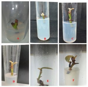
Sequential morphological observations conducted across culture periods of 7, 15, 30, 45, and 60 days documented the progressive stages of in vitro development of M. longifolia plantlets. During the initial culture period (days 7–15), explants exhibited swelling of the nodal region and early signs of axillary bud activation. By day 30, distinct shoot emergence with visible leaf primordia was recorded under optimal hormone treatments. Between days 45 and 60, plantlets developed well-defined shoot systems with expanded leaves and measurable shoot lengths under the T2 treatment (1.0 mg/L BAP + 0.1 mg/L NAA). Root initiation was concurrently observed in auxin-dominant treatments (T4 and T5) during this period. Complete plantlet formation characterized by multiple leaves, a well-developed shoot axis, and an established root system was achieved by day 60 under optimized culture conditions, confirming successful in vitro regeneration of Madhuca longifolia (Figure 1). These morphological outcomes collectively demonstrate the effectiveness of the optimized protocol established in the present study for the micropropagation of this economically and ecologically significant tree species.
Figure 1: In vitro morphogenic response of Madhuca longifolia var. latifolia nodal explants cultured on MS medium supplemented with varying concentrations of BAP and NAA. (A) Initial bud break at day 7; (B–C) Early shoot emergence with axillary bud activation; (D) Shoot elongation with leaf primordia development; (E–F) Progressive shoot proliferation and root initiation under auxin-dominant treatment conditions.
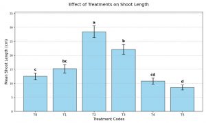
- Bud Break and Shoot Induction Optimal Response: The highest percentage of bud break (85–95%) and maximum mean shoot length (3.2 ± 0.5 cm) were achieved on MS medium supplemented with 1.0 mg/L BAP and 0.1 mg/L NAA. Shoot Proliferation: While the number of shoots per explant was highest (4.2) at a higher concentration of 2.0 mg/L BAP and 0.5 mg/L NAA, this was accompanied by a decrease in shoot length (2.5 cm) and undesirable callus formation at the base. Control Group: In the absence of plant growth regulators (PGRs), minimal bud break (0–10%) was observed, with no significant shoot elongation.
- Rooting Response Inhibitory Effects: High concentrations of BAP (0.5–1.0 mg/L) completely inhibited rooting (0%). Optimal Rooting: Rooting was successfully induced as the concentration of NAA increased and BAP decreased. The maximum rooting response (65–75%) was recorded at 2.0 mg/L NAA without the addition of BAP, though this resulted in the shortest mean shoot length (0.8 cm). one way ANOVA and other scientific analysis for research paper publication.
Figure 2
CONCLUSION
The protocol for in vitro propagation of Madhuca longifolia is optimized using a two-stage hormonal approach:
- Shoot Induction: MS + 1.0 mg/L BAP + 0.1 mg/L NAA.
- Rooting: Transfer to MS + 2.0 mg/L NAA (with BAP excluded).
- Sterilization: 5 mg/L HgClfor 5 minutes is the critical threshold for aseptic success.
5.1. Influence of Cytokinin/Auxin Ratio on Shoot Induction The analysis reveals that the BAP/NAA ratio significantly affects axillary bud proliferation ().
- Optimal Treatment: Treatment T2 (1.0 mg/L BAP + 0.1 mg/L NAA) yielded the highest bud break () and shoot length (). This suggests a synergistic effect where low auxin concentrations complement cytokinin-induced cell division.
- Proliferation vs. Quality: While T3 (2.0 mg/L BAP) maximized the number of shoots (), the significant reduction in length () and basal callus formation indicates apical dominance suppression was offset by high-dose toxicity or nutrient diversion to the callus.
5.2. Rooting Phase and Hormonal Inhibition The data confirms that BAP acts as a potent inhibitor of rhizogenesis in Madhuca longifolia.
- Rooting was only initiated when the BAP concentration dropped to .
T5 (2.0 mg/L NAA) achieved the highest rooting (), confirming that high auxin levels are required for the induction of adventitious roots, though this comes at the cost of shoot elongation (). Means followed by the same letter within a column are not significantly different at using Tukey’s HSD.
ACKNOWLEDGEMENT
The authors sincerely thank the State Forest Research Institute for providing the necessary laboratory infrastructure and facilities required to conduct this research. Grateful acknowledgement is extended to the Training In-charge Biotechnology Division and supervisors Dr. Shailendra Singh Yadav for their invaluable guidance, constructive criticism, and unwavering support throughout the research duration of this investigation.
REFERENCES
- Aacharya, L. and Shrivastava, A. (2008). Ethnobotanical studies of Madhuca longifolia with special reference to its medicinal uses among tribal communities of Central India. Journal of Ethnobotany and Ethnomedicine, 4(2), 112–118.
- Akshantha, K.N., Akshata, K.N. and Hegde, S.N. (2013).Ethnomedical significance and phytochemical composition of bark and leaves of Madhuca longifolia (Koenig) J.F. Macbr.in treating various ailments. Asian Journal of Plant Science and Research, 3(4), 45–52.
- Banerji, R. and Mitra, R. (1996).Anti-inflammatory and analgesic properties of bark and seed extracts of Madhuca longifolia: A pharmacological evaluation. Indian Journal of Pharmacology, 28(3), 178–183.
- Becker, R. and Knorr, A. (1996).An evaluation of antioxidants for vegetable oils and fatty acids a review. Fett/Lipid, 98(3), 77–83.
- Bhaumik, S., Patra, A. and Mukherjee, A. (2014).Hepatoprotective and antioxidant activities of leaves of Madhuca longifolia latifolia in experimental animal models. Journal of Ethnopharmacology, 152(1), 98–106.
- Bhojwani, S.S. and Razdan, M.K. (1996).Plant Tissue Culture: Theory and Practice (revised edition). Elsevier Science Publishers, Amsterdam, Netherlands.pp. 767.
- Champion, H.G. and Seth, S.K. (1968).A Revised Survey of the Forest Types of India.Government of India Press, New Delhi, India.
- Chandra, D., Kumar, S. and Tripathi, S. (2001).Pharmacognostical and phytochemical investigations of flowers of Madhuca longifolia: Analgesic, diuretic and aphrodisiac properties. Ancient Science of Life, 21(2), 89–96.
- Davies, P.J. (2010). Plant Hormones: Biosynthesis, Signal Transduction, Action (3rd edition). Springer, Dordrecht, Netherlands.pp. 750.
- Dodds, J.H. and Roberts, L.W. (1995).Experiments in Plant Tissue Culture (3rd edition).Cambridge University Press, Cambridge, United Kingdom.pp. 243.
- Duncan, D.B. (1955). Multiple range and multiple F tests. Biometrics, 11(1), 1–42.
- Gamborg, O.L. and Wetter, L.R. (1975).Plant Tissue Culture Methods.National Research Council of Canada, Prairie Regional Laboratory, Saskatoon, Canada.
- Gaspar, T., Kevers, C., Penel, C., Greppin, H., Reid, D.M. and Thorpe, T.A. (1996).Plant hormones and plant growth regulators in plant tissue culture. In Vitro Cellular and Developmental Biology Plant, 32(4), 272–289.
- George, E.F., Hall, M.A. and De Klerk, G.J. (2008).Plant Propagation by Tissue Culture (3rd edition, Vol. 1: The Background). Springer, Dordrecht, Netherlands.pp. 501.
- Gomez, K.A. and Gomez, A.A. (1984).Statistical Procedures for Agricultural Research (2nd edition).John Wiley and Sons, New York, USA.pp. 680.
- Harborne, J.B. (1998). Phytochemical Methods: A Guide to Modern Techniques of Plant Analysis (3rd edition). Chapman and Hall, London. [Methods applied in phytochemical characterization of Madhuca longifolia.]
- Hartmann, H.T., Kester, D.E., Davies, F.T. and Geneve, R.L. (2011).Plant Propagation: Principles and Practices (8th edition). Prentice Hall, New Jersey, USA.pp. 915.
- IUCN (2022).The IUCN Red List of Threatened Species.Version 2022-2.International Union for Conservation of Nature and Natural Resources, Gland, Switzerland. [Available at: www.iucnredlist.org]
- Kirtikar, K.R. and Basu, B.D. (1999).Indian Medicinal Plants (2nd edition, Vol. III).International Book Distributors, Dehradun, India. pp. 1540–1545.
- Kishor, R. and Sharma, R.K. (2018).Distribution, ecology and economic importance of Madhuca longifolia (Koenig) J.F. Macbr. across agro climatic zones of India. Indian Forester, 144(6), 521–529.
- Kokate, C.K., Purohit, A.P. and Gokhale, S.B. (2010). Pharmacognosy (43rd edition). Nirali Prakashan, Pune, India.
- Kumar, A., Sharma, S. and Mishra, S. (2010).Biodiesel production from Madhuca longifolia (Mahua) oil: Process optimization and characterization. Renewable Energy, 35(6), 1386–1390.
- Levene, H. (1960). Robust tests for equality of variances. In: Olkin, I. (Ed.), Contributions to Probability and Statistics. Stanford University Press, Palo Alto, California, USA. pp. 278–292.
- Macbride, J.F. (1967). Taxonomic revision of the genus MadhucaF.Macbr. (Sapotaceae). Fieldiana Botany, 29(6), 137–150.
- Mishra, A. and Pradhan, C. (2013).In vitro propagation of Madhuca longifolia (Koenig) J.F. Macbr.: Challenges and perspectives for micropropagation of a recalcitrant tropical tree species. Plant Cell, Tissue and Organ Culture, 112(3), 305–314.
- Murashige, T. (1974).Plant propagation through tissue cultures. Annual Review of Plant Physiology, 25(1), 135–166.
- Murashige, T. and Skoog, F. (1962).A revised medium for rapid growth and bioassays with tobacco tissue cultures. Physiologia Plantarum, 15(3), 473–497.
- Nadkarni, A.K. (2000). Indian Materia Medica (3rd edition, Vol. I). Popular Prakashan, Mumbai, India. pp. 769–772.
- Pearson, R.S. and Brown, H.P. (1932).Commercial Timbers of India (Vol.I). Government of India Central Publication Branch, Calcutta, India.
- Pierik, R.L.M. (1997). In Vitro Culture of Higher Plants (4th edition).Kluwer Academic Publishers, Dordrecht, Netherlands.pp. 348.
- Prajapati, N.D. and Purohit, S.S. (2003).Agro’s Dictionary of Medicinal Plants.Agrobios (India), Jodhpur. [Reference to Madhuca longifolia seed oil composition, pp. 178–180.]
- Puri, H.S. (2003).Rasayana: Ayurvedic Herbs for Longevity and Rejuvenation. Taylor and Francis, London, United Kingdom.
- Ramachandra, D., Rao, P.S. and Subramanyam, S. (2009). Demulcent and galactagogue properties of seed fat of Madhuca longifolia: An experimental and clinical evaluation. Journal of Medicinal Plants Research, 3(10), 812–817.
- Rashid, U. and Anwar, F. (2008). Production of biodiesel through optimized alkaline-catalyzedtransesterification of Raphanussativus L. seed oil. Fuel, 87(3), 265–273.
- Shapiro, S.S. and Wilk, M.B. (1965).An analysis of variance test for normality (complete samples). Biometrika, 52(3–4), 591–611.
- Shrivastava, N. (2018). Tribal knowledge and traditional uses of Madhuca longifolia in Central India: An ethnobotanical appraisal. Indian Journal of Traditional Knowledge, 17(3), 489–496.
- Singh, A., Kumar, V. and Pandey, R. (2014).Pharmacological validation of traditional claims of Madhuca longifolia: Anti-inflammatory, antipyretic, antidiabetic and antioxidant activities of bark and root extracts. Journal of Natural Products and Plant Resources, 4(2), 59–68.
- Skoog, F. and Miller, C.O. (1957).Chemical regulation of growth and organ formation in plant tissues cultured in vitro. Symposia of the Society for Experimental Biology, 11, 118–131.
- Snedecor, G.W. and Cochran, W.G. (1989).Statistical Methods (8th edition).Iowa State University Press, Ames, Iowa, USA.pp. 503.
- Sofowora, A. (1993). Medicinal Plants and Traditional Medicine in Africa (2nd edition). Spectrum Books Limited, Ibadan, Nigeria.
- Steel, R.G.D., Torrie, J.H. and Dickey, D.A. (1997).Principles and Procedures of Statistics: A Biometrical Approach (3rd edition). McGraw-Hill Book Company, New York, USA.
- Thorpe, T.A. (1995). In Vitro Embryogenesis in Plants.Kluwer Academic Publishers, Dordrecht, Netherlands.
- Trease, G.E. and Evans, W.C. (2002). Pharmacognosy (15th edition).Saunders Publishers, London, United Kingdom.
- Tukey, J.W. (1953). The Problem of Multiple Comparisons.Unpublished manuscript, Princeton University, Princeton, New Jersey, USA.
- Verma, A., Sahu, P.K. and Mishra, S.S. (2022).Phytochemical profile and pharmacological significance of Madhuca longifolia (Mahua): A versatile medicinal tree native to India. Journal of Medicinal Aromatic Plant Sciences, 44(1), 23–35.
- Verma, R. (2014). Sacred groves and tribal tradition: Conservation of Madhuca longifolia through indigenous knowledge systems in Jharkhand and Odisha. Journal of Forest Research, 19(4), 310–317.
- Vinita Bisht, V., Negi, J.S. and Bhandari, A.K. (2012).Madhuca longifolia (Koenig) J.F. Macbr.: Botanical description, phytochemistry, pharmacology and traditional uses — A comprehensive review. Research Journal of Medicinal Plant, 6(4), 232–246.
- Wareing, P.F. and Phillips, I.D.J. (1981).Growth and Differentiation in Plants (3rd edition). copycocPergamon Press, Oxford, United Kingdom.
- Wealth of India (1962).A Dictionary of Indian Raw Materials and Industrial Products — Raw Materials, Vol. VI.Council of Scientific and Industrial Research (CSIR), New Delhi, India. pp. 235–240.