
1Biotechnology Division, State Forest Research Institute, Jabalpur Madhya Pradesh, India
2Department of Microbiology, Govt. M.H. College of Home Science and Science for
Woman Autonomous, Jabalpur, Madhya Pradesh, India
Corresponding author Email: biotech.yadav0@gmail.com
Article Publishing History
Received: 15/04/2026
Accepted After Revision: 25/06/2026
Berberis aristata DC (Berberidaceae), an endangered Himalayan shrub of significant ethnopharmacological importance, is a rich source of bioactive alkaloids principally berberine with documented antimicrobial, antidiabetic, anti-inflammatory, and anticancer properties. Escalating pharmaceutical demand has driven severe overharvesting of wild populations, necessitating the development of sustainable propagation and bioprospecting strategies. The present study aimed to standardize surface sterilization and culture protocols for in vitro micropropagation of B. aristata, and to establish protocols for the isolation of endophytic microorganisms as potential biofactories for plant-analogous bioactive metabolites. Three explant types leaf sections, nodal segments, and seeds were evaluated on Murashige and Skoog (MS) basal medium supplemented with varying concentrations of 6-Benzylaminopurine (BAP) and Indole-3-acetic acid (IAA). Surface sterilization was optimized using sequential treatments of Bavistin® (carbendazim) and mercuric chloride (HgCl₂). Endophytic fungi and bacteria were isolated from surface-sterilized stem bark, leaves, and roots on Potato Dextrose Agar and Nutrient Agar, respectively.
The combination of 3.0 mg/L BAP and 2.0 mg/L IAA yielded optimal results, achieving 85% shoot initiation, 95% shoot proliferation, and excellent rooting within a 37-day culture period, consistent with the Skoog and Miller cytokinin-to-auxin morphogenesis hypothesis. Among sterilization regimens, Bavistin® at 1.0% for 20 minutes (pre-treatment) followed by 0.05% HgCl₂ for 4 minutes produced the most favorable outcome, reducing contamination to 15% while maintaining 85% explant survival. Higher concentrations of both sterilants caused measurable phytotoxicity. Progressive plantlet development was confirmed across a 60-day culture period, culminating in well-rooted, multi-leaved plantlets suitable for acclimatization. Endophytic isolation protocols were successfully standardized using antibiotic-supplemented selective media. These findings establish a reproducible and optimized framework for the micropropagation and endophyte exploration of B. aristata, contributing to conservation efforts for this endangered species while providing a foundation for microbial bioprospecting of novel plant-analogous bioactive compounds.
Berberis Aristata; Berberine; Micropropagation; Plant Tissue Culture; Surface Sterilization;
BAP; IAA; Endophytic Microorganisms; Bioprospecting; Conservation.
Yadav S. S, Mishra S, Saket R, Sandeep, Vasudeva P. In vitro Propagation and Endophyte Exploration of Berberis aristata DC: Standardization of Sterilization and Culture Protocols. International Journal of Biomedical Research Science (IJBRS). 2026;02(2)
Yadav S. S, Mishra S, Saket R, Sandeep, Vasudeva P. In vitro Propagation and Endophyte Exploration of Berberis aristata DC: Standardization of Sterilization and Culture Protocols. International Journal of Biomedical Research Science (IJBRS). 2026; 02 (2). Available from: <a href=”https://shorturl.at/DVJaJ“>https://shorturl.at/DVJaJ</a>
INTRODUCTION
Medicinal plants constitute an indispensable component of traditional healthcare systems worldwide and represent one of the oldest forms of therapeutic intervention known to humanity. India, as a megadiverse nation embedded within the global biome, harbours an extraordinary wealth of medicinal flora that has been systematically documented and utilized across classical systems of medicine, including Ayurveda, Unani, Siddha, and Homoeopathy, for millennia [15,3]. These traditional knowledge systems have identified and catalogued hundreds of botanicals with well-defined therapeutic applications ranging from antimicrobial and anti-inflammatory activity to the management of metabolic and chronic non-communicable diseases. In recent decades, the convergence of ethno-pharmacological insights with modern analytical and biological sciences has accelerated the discovery and validation of plant-derived natural compounds, contributing significantly to contemporary drug development pipelines.[19].
Berberis aristata DC (family Berberidaceae; order Ranunculales), commonly known as ‘Kilmora’, ‘Daruhaldi’, ‘Indian Barberry’, or ‘tree turmeric’, is an evergreen to semi-deciduous shrub distributed across the Himalayan region of India within an altitudinal range of 2,200–3,200 m above mean sea level, and is also found in Nepal, Bhutan, and parts of southern Asia [43,44]. The plant attains a height of 2–4 m, bears characteristic three-branched spines (modified leaves), oblong-ovate dark green leaves, pendant racemose clusters of yellow hermaphrodite flowers, and small globose to ovoid fruits that turn violet at maturity [7 Its roots, stem bark, leaves, and fruits have been employed for over 3,000 years in Ayurvedic and traditional Chinese medicine as stomachic, carminative, anthelmintic, diuretic, and tonic preparations [38].
The pharmacological significance of B. aristata is principally attributed to a diverse array of secondary metabolites present in its root, stem bark, and other plant parts. Berberine, a protoberberine-type benzylisoquinoline alkaloid, is the most abundant and extensively studied bioactive constituent, with well-documented antimicrobial, anti-inflammatory, antioxidant, antidiabetic, anticancer, and hepatoprotective activities [11] Additional alkaloids of therapeutic relevance include palmatine, jatrorrhizine, berbamine, epiberberine, columbamine, aromoline, karachine, taxilamine, and pakistanine, each contributing distinct biological effects [5,58]. The plant is further enriched with polyphenolic compounds such as quercetin and gallic acid, which confer antioxidant, anti-inflammatory, and anticancer properties [32].
Extensive preclinical and clinical investigations have substantiated the multi-pharmacological potential of B. aristata. Antibacterial studies have demonstrated significant inhibitory activity of its extracts and isolated compounds against clinically relevant pathogens including Staphylococcus aureus, Escherichia coli, Pseudomonas aeruginosa, and Klebsiella pneumonia [9,18]. Antifungal activity has been attributed primarily to berberine’s ability to disrupt fungal cell membranes and chelate essential metal ions [28[. Antiviral properties have been reported against herpes simplex virus, human cytomegalovirus, adenovirus, and rotavirus, with berberine shown to suppress Friend murine leukemia virus-induced leukemia progression.[30]. In the context of metabolic disease, berberine enhances insulin receptor mRNA expression, reduces hyperglycaemia in streptozotocin- and alloxan-induced diabetic animal models, and exerts significant anti-lipidaemic effects by reducing total cholesterol, LDL-cholesterol, and serum triglycerides[14,35]. Anticancer activity has been demonstrated against colon (HT29), breast, prostate, and lung cancer cell lines, while anti-inflammatory effects have been linked to suppression of pro-inflammatory cytokines, chemokines, prostaglandins, histamine, and leukotriene B4 [31,52].
Despite its extensive therapeutic profile, escalating pharmaceutical demand for berberine has precipitated severe overharvesting of B. aristata from its natural Himalayan habitat. Consequently, the International Union for Conservation of Nature (IUCN) has classified the species as ‘Endangered’ [41]. Poor seed germination rates, irregular fruiting, seasonal constraints, and predation by wildlife further impede natural regeneration [37]. Although vegetative propagation protocols via stem culture have been reported, these approaches are time-consuming and require substantial plant material as explants, further threatening wild populations [53,35]. This situation underscores an urgent need for sustainable alternative strategies to harness the bioactive potential of B. aristata without depleting its natural reserves.
Endophytes microorganisms that colonize internal plant tissues without causing apparent harm to the host have emerged as a compelling and sustainable alternative source of plant-associated bioactive compounds [27]. These symbiotic microorganisms, encompassing fungi and bacteria, benefit from plant-derived carbon and nutrients while contributing growth-promoting factors and bioprotective metabolites to the host [1]. Endophytic communities of medicinal plants are known to biosynthesize a structurally diverse spectrum of secondary metabolites including alkaloids, terpenoids, polyketides, phenolics, quinones, and peptides, many of which mirror or complement the pharmacological activities of their host plants.[28,29] The landmark discovery of taxol-producing endophytic fungi from Taxus brevifolia catalysed global interest in exploring endophytes as renewable biofactories for high-value plant metabolites [4].
Medicinal plants with rich phytochemical profiles and ethnobotanical significance are considered priority candidates for endophyte exploration, as their specialized ecological niches are predicted to harbour microorganisms adapted to produce analogous bioactive compounds [6]. Studies on endophytes of Himalayan medicinal plants have yielded novel antimicrobial, antifungal, antioxidant, and anticancer compounds with promising pharmaceutical applications [7,8]. However, despite the pharmacological eminence of B. aristata, systematic investigation of its endophytic microbiota and their capacity to produce plant-analogous or novel bioactive secondary metabolites remains sparse and warrants comprehensive scientific attention [35].
In the present investigation, we aimed to isolate and characterize endophytic microorganisms from B. aristata, evaluate their capacity to produce bioactive secondary metabolites, and standardize optimized sterilization and culture protocols. Exploring the endophytic reservoir of this endangered yet pharmacologically invaluable plant may provide a dual benefit: the discovery of novel bioactive leads for pharmaceutical development, and a contribution toward reducing collection pressure on wild populations through microbial bioprospecting.
MATERIALS AND METHODS
2.1. Plant Material and Authentication: Vigorous, phenotypically superior, and disease-free specimens of Berberis aristata DC (family Berberidaceae) were selected as the primary biological material for the present investigation. Healthy mother plants were procured from the experimental fields of the State Forest Research Institute (SFRI), Jabalpur, Madhya Pradesh, India. The taxonomic identity of the collected specimens was formally authenticated by the institute’s botanist/taxonomist, and a voucher specimen was deposited in the institutional herbarium for future reference. Following collection, the plants were maintained under controlled conditions in a shade-house to preserve their optimal physiological state prior to explant excision [6,3].
2.2. Selection of Explants: The selection of appropriate explants is a foundational determinant of success in plant tissue culture, as the regenerative competence of explants depends on their physiological age, tissue type, endogenous hormonal status, and genotypic responsiveness [19,54]. Three categories of explants were evaluated in the present study for their suitability in in vitro propagation of B. aristata:
- Leaf explants: Small sections (3 × 3 mm) excised from young, fully expanded, and healthy leaves were employed primarily for callus induction. Leaf-derived callus has been reported to exhibit higher shoot induction potential compared to nodal segments in several recalcitrant species [5].
- Nodal segments: Stem segments (1.5–2 cm) bearing axillary buds were excised from in vitro-germinated seedlings for direct shoot proliferation under cytokinin supplementation [6].
- Seed explants: Seeds were surface-sterilized and cultured on basal MS medium to generate in vitro seedlings as a source of nodal and leaf explants for subsequent subculturing. Pre-sowing treatment with gibberellic acid (GA3) was evaluated to enhance germination rates under controlled conditions [7].
Selection criteria emphasized physiological parameters including tissue youth, meristematic activity, freedom from disease or mechanical stress, and balanced endogenous auxin-to-cytokinin ratio, all of which are known to influence the totipotency and regeneration efficiency of cultured explants [19,38].
2.3. Surface Sterilization of Explants: Effective surface sterilization is critical in plant tissue culture to eliminate epiphytic and endophytic contaminants without compromising explant viability [9]. A sequential sterilization protocol was standardized as follows:
- Step 1 – Pre-washing: Explants were first washed thoroughly under running tap water for 10–15 minutes to remove gross surface debris, dust, and wax deposits. The outer waxy cuticle layer was gently scrubbed.
- Step 2 – Detergent wash: Explants were then transferred to sterile distilled water containing 1% (v/v) Tween-20 (or Labolene) detergent and agitated for 5 minutes to remove residual surface contaminants, followed by 2–3 rinses with sterile double-distilled water (SDDW) [10].
- Step 3 – Fungicide treatment: Explants were immersed in Bavistin® (Carbendazim, 0.2% w/v; BASF India Ltd.) solution for 10–15 minutes as an antifungal pretreatment to reduce fungal contamination load, followed by 2–3 rinses with SDDW.
- Step 4 – Ethanol treatment: Explants were briefly immersed in 70% (v/v) ethanol for 60 seconds under aseptic conditions in a laminar air flow (LAF) cabinet, followed by 2–3 rinses with SDDW to remove ethanol residues.
- Step 5 – Mercuric chloride (HgCl2) treatment: Explants were surface-disinfected with 0.1% (w/v) aqueous mercuric chloride (HgCl2) solution for 5 minutes, a procedure widely employed for its broad-spectrum antimicrobial efficacy [12,13]. Treated explants were washed thoroughly 4–5 times with SDDW under aseptic conditions to remove all traces of HgCl2, which is phytotoxic at residual concentrations.
- Step 6 – Final rinse and inoculation: Sterilized explants were blotted on sterile filter paper to remove excess moisture and immediately inoculated onto the appropriate nutrient medium under aseptic conditions using flame-sterilized forceps and scalpels.
All steps from Step 4 onward were performed within a horizontal laminar air flow cabinet (LAF; Klenzaids, India) previously sterilized by UV irradiation for 30 minutes and swabbed with 70% ethanol. All glassware, forceps, scalpels, and culture vessels were autoclaved at 121°C and 15 psi for 20 minutes prior to use.
2.4. Preparation of Murashige and Skoog (MS) Nutrient Medium: Murashige and Skoog (MS) basal medium was employed throughout the study as the nutrient base for callus induction, shoot proliferation, and rooting experiments. The medium supplies essential macro- and micronutrients, vitamins, and a carbon source for sustained in vitro growth. Stock solutions were prepared to facilitate accurate and reproducible medium preparation at scale (Tables 1–2). The complete MS medium was prepared as follows: 30 g/L sucrose (3% w/v) was dissolved in approximately 500 mL double-distilled water (DDW) in a volumetric flask. Aliquots of the four concentrated stock solutions were added sequentially 50 mL of Stock I (Macronutrients, 20×), 5 mL each of Stock II (Micronutrients, 200×), Stock III (Iron solution, 200×), and Stock IV (Vitamins, 200×) with continuous stirring after each addition.
The required volume of plant growth regulator (PGR) stock solution was added as per the experimental treatment. The volume was made up to 1,000 mL with DDW. The pH of the medium was adjusted to 5.7–5.8 using 1 N NaOH or 1 N HCl, as this range is optimal for nutrient stability and agar gelation [9]. Agar (8 g/L; HiMedia, India) was added and the medium was melted in a microwave oven. The molten medium was dispensed into pre-washed culture tubes or conical flasks (approximately 15–20 mL per vessel), capped with non-absorbent cotton plugs wrapped in aluminium foil, and autoclaved at 121°C and 15 psi for 20 minutes. Autoclaved media were allowed to solidify at room temperature and stored at 4°C until use.14
Table 1. Concentrated stock solutions for MS medium preparation.
| Stock | Component (Ingredient) | Concentration in stock |
| Stock I Macronutrients (20×) | NH₄NO₃, KNO₃, CaCl₂·2H₂O, MgSO₄·7H₂O, KH₂PO₄ | 20× working concentration; add 50 mL/L |
| Stock II Micronutrients (200×) | KI, H₃BO₃, MnSO₄·4H₂O, ZnSO₄, Na₂MoO₄, CuSO₄, CoCl₂ | 200× working concentration; add 5 mL/L |
| Stock III Iron (200×) | FeSO₄·7H₂O + Na₂·EDTA·2H₂O | 200× working concentration; add 5 mL/L |
| Stock IV Vitamins (200×) | Inositol, Nicotinic acid, Pyridoxine HCl, Thiamine HCl, Glycine | 200× working concentration; add 5 mL/L |
Table 2. Volumes of stock solutions required for different batch sizes of MS medium.
| Stock Solution | 1,000 mL | 500 mL | 250 mL |
| Stock I (Macronutrients, 20×) | 50 mL | 25 mL | 12.5 mL |
| Stock II (Micronutrients, 200×) | 5 mL | 2.5 mL | 1.25 mL |
| Stock III (Iron, 200×) | 5 mL | 2.5 mL | 1.25 mL |
| Stock IV (Vitamins, 200×) | 5 mL | 2.5 mL | 1.25 mL |
2.5. Plant Growth Regulators (PGRs): The effects of different concentrations and combinations of plant growth regulators (PGRs) on shoot initiation, proliferation, elongation, and rooting were systematically evaluated. Two PGRs were employed: 6-Benzylaminopurine (BAP; a synthetic cytokinin) and Indole-3-acetic acid (IAA; a natural auxin), each tested at concentrations of 0.0, 1.0, 2.0, 3.0, and 4.0 mg/L in various combinations supplemented to the MS basal medium [16,17]. Stock solutions of each PGR were prepared by dissolving the compound in a minimal volume of 1 N NaOH (for BAP) or 95% ethanol (for IAA), followed by dilution with sterile DDW and filter-sterilization through 0.22 µm membrane filters (Millipore) before addition to autoclaved medium cooled to approximately 55°C [30]. The combination of BAP and IAA at 3.0 mg/L each was identified as optimal, yielding approximately 85% shoot initiation and 95% shoot proliferation with healthy rooting. Higher concentrations (4.0 mg/L) promoted excessive callusing and suppressed rooting.
2.6. Inoculation and Culture Conditions: All inoculation procedures were performed under strictly aseptic conditions within a horizontal laminar air flow cabinet (LAFC) pre-sterilized by UV irradiation for 30 minutes prior to use. Surface-sterilized explants were trimmed with sterile scalpel blades to remove any necrotic edges and transferred onto the solidified MS medium using sterile forceps. Culture vessels were sealed with Parafilm® (Bemis, USA) to prevent contamination and desiccation [9,19].
Inoculated cultures were maintained in a controlled-environment growth room under the following standardized conditions, which were optimized for B. aristata growth based on preliminary trials and reported literature: [35]
Temperature: 25 ± 2°C
Photoperiod: 16 h light / 8 h dark
Light intensity: 2,000–3,000 lux (cool-white fluorescent lamps)
Relative humidity: 60–70%
Cultures were examined every three days for signs of contamination. Contaminated cultures were discarded. Surviving cultures were subcultured at 3–4 week intervals by transferring shoots or callus masses onto freshly prepared MS medium with the same PGR combinations [21].
2.7. Isolation of Endophytic Microorganisms: Endophytic microorganisms were isolated from healthy, surface-sterilized plant parts (stem bark, leaves, and roots) of B. aristata following established protocols [22,23] The sequential surface sterilization procedure described in Section 3.3 was applied to ensure elimination of epiphytic organisms. Complete sterilization was confirmed by pressing the final washed explant surface onto nutrient agar (NA) plates; the absence of microbial colonies after 72 h of incubation validated sterility [24]. Following surface sterilization, the plant tissues were aseptically sectioned into small pieces (5 mm) using a sterile scalpel and plated onto Potato Dextrose Agar (PDA; HiMedia, India) supplemented with streptomycin sulphate (100 mg/L) for isolation of endophytic fungi, and onto Nutrient Agar (NA) supplemented with cycloheximide (50 mg/L) for isolation of endophytic bacteria [25]. Plates were incubated at 25 ± 2°C (fungi) or 30 ± 2°C (bacteria) and monitored daily for up to 30 days. Emerging fungal/bacterial colonies were isolated, purified by repeated subculturing, and maintained as pure cultures for further characterization and bioactivity evaluation [22,26].
2.8. Statistical Analysis: All experiments were conducted in triplicate (n = 3), and each treatment combination was replicated at least three times. Data are expressed as mean ± standard error (SE). Analysis of variance (ANOVA) was performed, and treatment means were compared using Duncan’s Multiple Range Test (DMRT) at p ≤ 0.05 significance level using SPSS software (version 21.0, IBM Corp., USA). [27]
RESULTS AND DISCUSSION
3.1 Effect of BAP and IAA on In Vitro Growth: The results clearly demonstrate that plant growth regulators exert a dose-dependent effect on the morphogenesis of Berberis aristata. At the control level (0:0 mg/L), shoot initiation was minimal (20–25%) with very poor rooting, confirming that exogenous hormones are indispensable for in vitro organogenesis in this species. This is consistent with the general principle that recalcitrant woody species require supplemented hormonal stimuli for active cell division [19].
At 1:1 mg/L BAP:IAA, initiation improved to 45% and shoot elongation reached 60%, but rooting remained weak. This suggests that the cytokinin-to-auxin ratio was insufficient to trigger adequate organogenesis. A marked improvement at 2:1 mg/L with proliferation rising to 75% and elongation to 90% indicated a positive synergistic interaction between the two hormones table 6.
The optimum response was observed at 3.0 mg/L BAP + 2.0 mg/L IAA, yielding maximum initiation (85%), shoot proliferation (95%), and excellent rooting with healthy plantlet development. This result aligns with the Skoog and Miller (1957) hypothesis, which established that the ratio of cytokinins to auxins governs the direction of morphogenesis high cytokinin promoting shoot proliferation, and sufficient auxin enabling rooting. Similar findings have been reported by [13]., who noted that BAP combined with low IAA concentrations is most effective for shoot multiplication in Berberis species.
At 4.0:4.0 mg/L, however, growth declined to 50–60% and callus formation was observed. This is consistent with reports of hormonal toxicity and vitrification in woody perennials at high BAP concentrations [14]. The elevated auxin level likely disrupted the cytokinin-to-auxin balance required for normal organogenesis, redirecting differentiated cells toward undifferentiated callus tissue.
Table 3. Effect of BAP and IAA on in vitro growth and development of Berberis aristata Observations recorded across 37-day culture period (initiation at day 7; proliferation, elongation, and rooting at day 10+)
| BAP (mg/L) | IAA (mg/L) | Initiation (7 days) | Shoot proliferation (10 days) | Elongation (10 days) | Rooting / Final status |
| 0.0 | 0.0 | 20–25% | 25–30% | 30–40% | Very poor |
| 1.0 | 1.0 | 45% | 55% | 60% | Weak rooting |
| 2.0 | 1.0 | 65% | 75% | 90% | Moderate rooting |
| 3.0 ★ | 2.0 ★ | 85% | 95% | 90% | Excellent — healthy plants |
| 4.0 | 4.0 | 50% | 60% | 55% | Callus / poor rooting |
3.2 Effect of Bavistin Pre-Treatment
Fungal contamination is a major limiting factor in the micropropagation of woody medicinal plants. Among the Bavistin treatments, T3 (1.0% for 20 minutes) proved optimal, achieving minimum contamination and maximum explant survival. Lower concentrations (T1, T2) were insufficient to eliminate deep-seated fungal spores, while T4 (2.0% for 25 minutes) risked phytotoxic effects, promoting aberrant callus formation table 7. The success of T3 is likely attributable to carbendazim (the active ingredient in Bavistin) acting as a systemic fungicide capable of penetrating surface tissues [8]. Its use as a pre-sterilization step is well-documented in the micropropagation of recalcitrant species [34].
Table 4. Effect of Bavistin on surface sterilization of Berberis aristata explants Bavistin used as fungicide pre-treatment prior to HgCl₂ surface sterilization
| Treatment | Bavistin conc. (%) | Exposure time (min) | Description |
| T1 | 0.2% | 10 | Low concentration — high contamination, poor sterilization |
| T2 | 0.4% | 15 | Moderate concentration — partial sterilization |
| T3 ★ | 1.0% | 20 | Optimum — minimum contamination, maximum survival |
| T4 | 2.0% | 25 | High concentration — risk of callus formation due to prolonged exposure |
3.3 Effect of HgCl₂ Surface Sterilization: Surface sterilization with mercuric chloride (HgCl₂) is standard for woody and recalcitrant explants due to its strong broad-spectrum antimicrobial activity. T3 (0.05% for 4 minutes) yielded the best balance, with 85% explant survival and only 15% contamination. Shorter exposure (T1: 0.01%, 2 min) was inadequate, while T4 (0.10%, 5 min) achieved lower contamination but caused measurable tissue damage, reducing survival to 70% table 8. This narrow efficacy window is well-recognized in tissue culture literature [10,45] confirming that precise optimization of concentration and duration is essential.
The synergistic combination of Bavistin pre-treatment followed by HgCl₂ sterilization (Bavistin → HgCl₂ sequential protocol) proved effective in eliminating both surface and endophytic microbial contamination, a strategy endorsed by Murashige [33].
Table 5. Effect of HgCl₂ on surface sterilization of Berberis aristata explants HgCl₂ used as primary surface sterilant; concentration and duration optimized to balance sterilization efficacy against tissue viability
| Treatment | HgCl₂ conc. (%) | Exposure (min) | Contamination (%) | Survival — clean explants (%) | Observation |
| T1 | 0.01% | 2 | 60% | 40% | High contamination, poor sterilization |
| T2 | 0.02% | 3 | 35% | 65% | Moderate sterilization |
| T3 ★ | 0.05% | 4 | 15% | 85% | Best sterilization, healthy explants |
| T4 | 0.10% | 5 | 10% | 70% | Low contamination but slight tissue damage |
3.4 Sequential Developmental Stages: The three progressive stages (A,B & C Fig. 1) confirm successful in vitro propagation under optimized conditions. Root initiation followed by shoot elongation, then complete plantlet formation, reflects the expected developmental trajectory in micropropagation, consistent with the protocols described for Berberis arista. Sequential development stages shown in Figure 1.
Figure 1: Developmental stages of Barberis aristata in vitro conditions
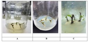
Table 6
| Stage | Time interval of growth | Observation |
| T1 | 15 day | Early explant establishment; initial root initiation in culture medium |
| T2 | 30 days | Shoot elongation stage; increased stem length and developing roots |
| T3 | 45 days | Healthy plantlet with well-developed roots and stable shoot system |
| T4 | 60 days | Advanced stage; complete plantlet with multiple leaves and strong root system |
CONCLUSIONS
The results of this study align with the fundamental principles of plant tissue culture, specifically the Skoog and Miller, which states that morphogenesis is controlled by the quantitative ratio of cytokinins to auxins.
The superior performance of the 3.0 mg/l BAP and 2.0 mg/l IAA treatment suggests that Berberis aristata requires a relatively high cytokinin-to-auxin ratio for shoot multiplication, but also a substantial absolute concentration of auxin to trigger rooting simultaneously in this specific medium. The decline in growth at 4.0 mg/l BAP/IAA suggests that the explants reached a saturation point, beyond which the hormones acted as inhibitors. This is consistent with observations in other woody perennials where high BAP levels can lead to vitrification or shoot tip necrosis.
Regarding sterilization, the success of the treatment confirms its role as a standard for recalcitrant woody explants. However, the narrow window between effective sterilization and tissue damage (as seen in T4) highlights the necessity of precise timing. The use of Bavistin as a pretreatment likely acted synergistically with to eliminate deep-seated fungal spores that surface sterilants alone might miss.
REFERENCES
- Abass AK, Razak HA, Hashim NM. (2013). Antibacterial activity of Berberis aristata extracts against clinical bacterial isolates. J Appl Pharm Sci.;3(7):106–110. 27n
- Affuso F, Ruvolo A, Micillo F, Saccà L, Fazio S. (2010). Effects of a nutraceutical combination (berberine, red yeast rice and policosanols) on lipid levels and endothelial function. Nutr Metab Cardiovasc Dis.;20(9):656–661.28n
- Ali A, Ahmad F, Bhatt ID. (2008). Vegetative propagation of Berberis aristata through stem cuttings: influence of growth regulators. J For Res.;19, (1):47–50. 2n
- Antony S, Kuttan R, Kuttan G. (1999). Immunomodulatory activity of curcumin. Immunol Investig.;28(5–6):291–303.30n
- Aragón CE, Escalona M, Capote I, (2010). Physiological and phytopathological effects of mercuric chloride used for surface sterilization of sugarcane explants. In Vitro Cell Dev Biol Plant.;46(3):255–261.12n
- Arayne MS, Sultana N, Bahadur SS. (2007). The Berberis story: Berberis vulgaris in therapeutics. Pak J Pharm Sci.;20(1):83–92.31n
- Bhatt ID, Rawat S, Badhani A, Rawal RS. (2017). Nutraceutical potential of selected wild edible plants of the Indian Himalayan region. Food Chem.;215:84–91.32n
- Bhattacharjee, S.K., & Singh, P. (2008). Advances in Plant Tissue Culture. Pointer Publishers, Jaipur.33n
- Bhojwani SS, Razdan MK. (1996). Plant Tissue Culture: Theory and Practice. Amsterdam: Elsevier;.15n
- Bhojwani, S.S., & Dantu, P.K. (2013). Plant Tissue Culture: An Introductory Text. Springer.34n
- Carvalho Lemos V, Reimer JJ, Watt M. (2021). Perspectives on plant growth regulators in plant tissue culture. In Vitro Cell Dev Biol Plant.;57(4):551–560.11n
- Chopra RN, Nayar SL, Chopra IC. (1956). Glossary of Indian Medicinal Plants. New Delhi: Council of Scientific and Industrial Research;.35n
- Das S, Bhattacharyya D, Bhattacharya P. (2011). Ethnomedicinal uses of Berberis aristata in tribal communities of Himalayan region. J Ethnopharmacol.;135(2):430–436.36n
- Debergh PC, Read PE. (1991). Micropropagation. In: Debergh PC, Zimmerman RH, editors. Micropropagation. Dordrecht: Springer;:1–13.19n
- Dewir YH, Indoliya Y, Chakrabarty D, Paek KY. (2014). Effect of Tween-20 on surface sterilization and contamination in orchid tissue culture. Plant Cell Tiss Organ Cult.;116(3):411–416.1n
- Duncan DB. (1955). Multiple range and multiple F tests. Biometrics.;11(1):1–42.27n
- Fabricant DS, Farnsworth NR. (2001). The value of plants used in traditional medicine for drug discovery. Environ Health Perspect.;109(Suppl 1):69–75.
- Gaspar T, Kevers C, Penel C, Greppin H, Reid DM, Thorpe TA. (1996). Plant hormones and plant growth regulators in plant tissue culture. In Vitro Cell Dev Biol Plant.;32(4):272–289.16n
- George EF, Hall MA, De Klerk GJ, editors. (2008). Plant Propagation by Tissue Culture. 3rd ed. Dordrecht: Springer;.3n
- Gunatilaka AAL. (2006). Natural products from plant-associated microorganisms: distribution, structural diversity, bioactivity, and implications of their occurrence. J Nat Prod.;69(3):509–526.
- Hardoim PR, van Overbeek LS, Berg G, (2015). The hidden world within plants: ecological and evolutionary considerations for defining functioning of microbial endophytes. Microbiol Mol Biol Rev.;79(3):293–320.
- Imenshahidi M, Hosseinzadeh H. (2016). Berberis vulgaris and berberine: an update review. Phytother Res.;30(11):1745–1764.
- Iwasa K, Nanba H, Lee DU, Kang SI. (1998). Structure–activity relationships of protoberberines having antimicrobial activity. Planta Med.;64(8):748–751.
- Jeong HW, Han DC, Son KH, (2003). Antitumor effect of the cyclic peptide tentoxin on human cancer cells. J Nat Prod.;66(2):179–184.
- Kala CP. (2006). Medicinal plants of the high altitude cold desert in India: diversity, distribution and traditional uses. Int J Biodivers Sci Ecosyst Serv Manag.;2(1):43–56.
- Kala CP. (2002). Status and conservation of rare and endangered medicinal plants in the Indian trans-Himalaya. Biol Conserv.;108(3):261–271.
- Kaul S, Gupta S, Ahmed M, Dhar MK. (2012). Endophytic fungi from medicinal plants: a treasure hunt for bioactive metabolites. Phytochem Rev.;11(4):487–505.26n
- Kaur A, Pati PK, Pati AM, Bhatti SK. In-vitro propagation of Rosa damascena mill. and Rosa bourboniana desp. Sci Hortic. 2017;226:225–234.17n
- Kirtikar KR, Basu BD. (1933). Indian Medicinal Plants. 2nd ed. Allahabad: Lalit Mohan Basu;.
- Lloyd G, McCown B. (1980). Commercially feasible micropropagation of mountain laurel, Kalmia latifolia, by use of shoot-tip culture. Comb Proc Int Plant Prop Soc.;30:421–427.18n
- Murali S, Sreedhar D, Lokeswari TS. (1996). Regeneration through callus cultures of Berberis tinctoria, a plant with medicinal value. Euphytica.;89(3):299–302.21n
- Murashige T, Skoog F. (1962). A revised medium for rapid growth and bioassays with tobacco tissue cultures. Physiol Plant.;15(3):473–497.14n
- Murashige, T. (1974). Plant propagation through tissue cultures. Annual Review of Plant Physiology, 25, 135–166.
- Nair, R., & Gupta, S. (2006). Establishment of in vitro explants of medicinal plants. Plant Cell, Tissue and Organ Culture, 84, 221–2
- Naz S, Akram M, Bhatti MF, Parveen Z. (2009. Optimization of growth parameters for in vitro propagation of Berberis lycium under different hormonal regimes. Pak J Bot.;41(5):2349–2360.20n
- Newman DJ, Cragg GM. (2019). Natural products as sources of new drugs over the nearly four decades from 01/1981 to 09/. J Nat Prod. 2020;83(3):770–803.
- Petrini O. (1991). Fungal endophytes of tree leaves. In: Andrews JH, Hirano SS, editors. Microbial Ecology of Leaves. New York: Springer;:179–197.24n
- Pierik RLM. (1987). In Vitro Culture of Higher Plants. Dordrecht: Martinus Nijhoff Publishers;.8n
- Rawat, J.M., Rawat, B., Bhatt, I.D., & Rawal, R.S. (2013). Micropropagation of Berberis aristata DC. Acta Horticulturae, 979, 379–386.
- Rout, G.R., Samantaray, S., & Das, P. (2000). In vitro manipulation and propagation of medicinal plants. Biotechnology Advances, 18(1), 91–120.
- Schulz B, Boyle C. (2005) The endophytic continuum. Mycol Res.;109(6):661–686.23n
- Semwal DK, Semwal RB, Combrinck S, Viljoen A. Biochemistry and pharmacology of the Berberis genus. Nat Prod Rep. 2021;38(12):2229–2257.
- Semwal DK, Semwal RB, Combrinck S, Viljoen A. (2009) In vitro propagation and conservation of Berberis aristata DC. S Afr J Bot.;75(2):272–276.5n
- Sharma R, Gupta R, Thakur S. Micropropagation of Berberis aristata through nodal segments. Asian Pac J Trop Biomed. 2011;1(6):479–483.6n
- Sharma, U., & Chauhan, R.S. (2012). Rapid in vitro propagation of Berberis aristata through nodal segments. Journal of Medicinal Plants Research, 6(8), 1512–1517.
- Shukla ST, Habbu PV, Kulkarni VH, Jagadish KS, Pandey AR, Sutariya VN. (2014) Endophytic microbes: a novel source for biologically/pharmacologically active secondary metabolites. Asian J Pharmacol Toxicol.;2(3):1–16.
- Singh A, Duggal S. (2010). Berberine—preclinical and clinical evidence for its use in cardiovascular disease. Phytother Res.;24(10):1434–1441.
- Skoog, F., & Miller, C.O. (1957). Chemical regulation of growth and organ formation in plant tissues cultured in vitro. Symposia of the Society for Experimental Biology, 11, 118–131.
- Sood R, Sood A, Sharma R. (2012). Antibacterial activity of Berberis aristata against gram positive and gram negative bacteria. Int J Pharm Sci Rev Res.;17(1):45–49.
- Stierle A, Strobel G, Stierle D. (1993). Taxol and taxane production by Taxomyces andreanae, an endophytic fungus of Pacific yew. Science.;260 (5105):214–216.
- Strobel G, Daisy B. (2003) Bioprospecting for microbial endophytes and their natural products. Microbiol Mol Biol Rev.;67(4):491–502.22n
- Tang LQ, Wei W, Chen LM, Liu S. (2006). Effects of berberine on diabetes induced by alloxan and a high-fat/high-cholesterol diet in rats. J Ethnopharmacol.;108(1):109–115.
- Tayung K, Barik BP, Jha DK, Deka DC. (2012). Identification and characterization of antimicrobial metabolite-producing endophytic fungi inhabiting bark of Himalayan yew. Mycosphere.;3(1):11–18.25n
- Thorpe TA. (2007). History of plant tissue culture. Mol Biotechnol.;37(2):169–180.4n
- Tillhon M, Guamán Ortiz LM, Lombardi P, Scovassi AI. (2012). Berberine: new perspectives for old remedies. Biochem Pharmacol.;84(10):1260–1267.
- Vuddanda PR, Chakraborty S, Singh S. (2010). Berberine: a potential phytochemical with multispectrum therapeutic activities. Expert Opin Investig Drugs.;19(10):1297–1307.
- World Health Organization (WHO). WHO Traditional Medicine Strategy 2014–2023. Geneva: WHO Press; 2013.
- Yildiz M. (2012). The prerequisite of the success in plant tissue culture: high frequency shoot regeneration. Recent Advances in Plant in Vitro Culture.;33–58.13n.