1Biotechnology Division, State Forest Research Institute, Jabalpur Madhya Pradesh, India
2Department of Microbiology, Govt. M.H. College of Home Science and Science for
Woman Autonomous, Jabalpur, Madhya Pradesh, India
Corresponding author Email: biotech.yadav0@gmail.com
Article Publishing History
Received:
Accepted After Revision:
Gmelina arborea Roxb. (family Lamiaceae), commonly known as Gamhar or Khmer, is a fast-growing multipurpose timber tree of significant ecological and commercial importance in tropical and subtropical forestry. Conventional propagation methods are inadequate for large-scale, genetically uniform planting stock production. The present study describes the development of an efficient in vitro micropropagation protocol for G. arborea using nodal stem segments from semi-hardwood (green-brownish) culm-stage stock plants maintained at the State Forest Research Institute (SFRI), Jabalpur, Madhya Pradesh, India. A sequential surface sterilization regime comprising 0.1% Bavistin (Carbendazim) with Tween-20 for 10–20 min, followed by 0.1% HgCl2 for 2 min, yielded optimal decontamination with minimal phytotoxicity.
Murashige and Skoog (MS) basal medium supplemented with varying concentrations of 6-Benzylaminopurine (BAP) and α-Naphthaleneacetic acid (NAA) served as the primary culture system. Culture conditions were maintained at 25 ± 2°C under a 16-hour photoperiod with white fluorescent LED illumination. Shoot bud induction, callus formation, and root initiation were evaluated over 4–8 week culture intervals. Experiments were arranged in a Completely Randomized Design (CRD), and treatment means were statistically separated by Duncan’s Multiple Range Test (DMRT) at p ≤ 0.05. Results indicate that elevated cytokinin-to-auxin ratios in MS medium significantly promoted axillary shoot proliferation, whereas high auxin concentrations favoured rhizogenesis. This protocol provides a reproducible, scalable framework for mass clonal propagation of elite G. arborea genotypes for afforestation and agroforestry programs.
Gmelina arborea, Micropropagation, Nodal Explants, MS Medium, BAP, NAA, Plant Tissue Culture, In vitro Propagation.
Yadav S. S, Upadhyay J, Dubey S, Sandeep, Vasudeva P. In vitro Propagation of Gmelina arborea Roxb. (Gamhar) Through Nodal Explants and Optimization of Sterilization and Culture Medium Protocols for Enhanced Micropropagation. International Journal of Biomedical Research Science (IJBRS). 2026; 2(2)
Yadav S. S, Upadhyay J, Dubey S, Sandeep, Vasudeva P. In vitro Propagation of Gmelina arborea Roxb. (Gamhar) Through Nodal Explants and Optimization of Sterilization and Culture Medium Protocols for Enhanced Micropropagation. International Journal of Biomedical Research Science (IJBRS). 2026; 2 (2). Available from: <a href=”https://shorturl.at/u7KsW“>https://shorturl.at/u7KsW</a>
INTRODUCTION
General Introduction: Gmelina arborea Roxb. (Family: Lamiaceae), commonly known as ‘Gamhar’ in Hindi and ‘White Teak’ in English, is a fast-growing deciduous tree that holds significant importance in tropical and subtropical forestry. It occurs naturally throughout the greater part of India at altitudes ranging from sea level up to 1500 meters (approximately 5000 ft), and has been extensively introduced as a fast-growing timber species since the 1960s.[3, 5, 10, 25]
The species is widely distributed across South and Southeast Asia, with its natural range encompassing India, Nepal, Bangladesh, Myanmar, Thailand, Laos, Cambodia, and Vietnam. Within India, it is found across several states including Madhya Pradesh, Uttar Pradesh, Bihar, Jharkhand, Chhattisgarh, Odisha, and Maharashtra.[16, 24, 26] The tree thrives in tropical and subtropical climates, preferring warm temperatures between 20–30°C, and grows best in moist fertile valleys with an annual rainfall ranging from 750 to 4500 mm.[10, 25]
G. arborea exhibits a preference for loamy and sandy loam soils and is commonly found in forests, riverbanks, and agricultural lands. It does not thrive on poorly drained soils and remains stunted on dry, sandy, or nutrient-deficient soils; drought conditions may further reduce it to a shrubby form. The species does not tolerate waterlogging, which restricts its cultivation to well-drained sites.[3, 10, 12]
1.2 Botanical Description and Scientific Classification: Botanically classified under the Kingdom Plantae, Division Magnoliophyta, Class Magnoliopsida, Order Lamiales, and Family Lamiaceae, Gmelina arborea is the type species of the genus Gmelina. The tree attains a moderate to large height of up to 30 m, with a girth of 1.2 to 4 m. Its bark is characteristically brown externally, with a chlorophyll layer just under the outer bark, and white on the inside.[7, 19, 26]
The wood of G. arborea is pale yellow to cream-coloured or pinkish-buff when freshly sawn, turning yellowish-brown upon exposure. It is classified as soft to moderately hard, light to moderately heavy, and lustrous, with a grain that is usually straight to irregular or rarely wavy, and a medium to coarse texture.[17, 19] Flowering occurs from February to April when the tree is more or less leafless, while fruiting commences from May through June. The fruit measures up to 2.5 cm in length, is smooth, dark green turning yellow when ripe, and emits a characteristic fruity smell.[7, 25]
1.3 Economic and Timber Importance: arborea is considered one of the most important species for commercial timber production in tropical Asia.[15, 26] Its timber is reasonably strong for its weight and is widely utilized in construction, furniture making, carriage manufacturing, sports equipment, musical instruments, and prosthetic (artificial) limbs. Once seasoned, the wood is very stable and moderately resistant to termites.[7, 19, 27]
It is highly esteemed in the woodworking industry for the manufacture of doors, windows, pencils, joinery, and furniture particularly for drawers, wardrobes, cupboards, kitchen cabinets, and other household furniture owing to its lightweight nature, dimensional stability, and durability.[4, 17, 26] Of considerable historical and cultural significance, the lion throne the most important and last surviving of the eight royal thrones of Myanmar now preserved at the National Museum in Yangon is carved from Gmelina arborea wood.[26]
1.4 Traditional and Medicinal Uses: In addition to its timber value, G. arborea has a long history of use in traditional medicine across South and Southeast Asia, particularly in Ayurveda and Khmer traditional medicine.[6, 11, 20] The bark and root preparations are employed to improve digestion and treat various stomach disorders. Decoctions of the roots are used as antipyretic agents to reduce fever. The plant also possesses anti-inflammatory properties, making it useful in alleviating joint swelling and pain. Furthermore, preparations from various plant parts are traditionally applied in the management of urinary tract disorders.[6, 11, 20]
Several pharmacological studies have corroborated these ethnomedicinal claims, reporting the presence of biologically active compounds such as alkaloids, flavonoids, saponins, and terpenoids in different parts of the plant.[18, 26]
1.5 Significance of the Present Study: Given its versatile utility spanning timber production, agroforestry, soil conservation, and traditional medicine, Gmelina arborea has attracted increasing scientific attention worldwide.[2, 16, 26] Despite its widespread use, systematic documentation of its morphological, anatomical, phytochemical, and pharmacological properties remains limited in the published literature.[18] The present study, therefore, aims to provide a comprehensive review of the botany, geographical distribution, timber characteristics, and medicinal properties of G. arborea, along with a synthesis of relevant scientific findings, contributing to the body of knowledge for future research and sustainable utilization of this valuable species.[26]
MATERIALS AND METHODS
2.1 Plant Material and Explants Selection: Healthy, disease-free stock plants of Gmelina arborea Roxb. (Khmer/Gamhar) were collected from the plantations maintained at the State Forest Research Institute (SFRI), Jabalpur, Madhya Pradesh, India. Nodal stem segments at the green-brownish (semi-hardwood) culm stage were selected as explants, as this developmental stage offers an optimal balance between meristematic competence and microbial manageability.[2, 8] The internodal regions of the stem were excised into segments of approximately 7–8 cm in length using a clean, sterile surgical blade. Only visually healthy, firm, and blemish-free nodes were selected to maximize regeneration potential.[8, 22]
2.2 Sterilization Protocols
2.2.1 Sterilization of Glassware and Equipment: All glassware including test tubes, culture bottles, conical flasks, measuring cylinders, pipettes, and Petri dishes were first thoroughly washed with a laboratory-grade detergent, rinsed with tap water, and subsequently with distilled water to remove all residual detergent. The glassware was then oven-dried at 60°C. Moist-heat sterilization was carried out using an autoclave at 121°C and 15 psi for 20–40 minutes (Table 1). Dry heat sterilization (160°–180°C for 1 hour) was applied to empty glassware such as pipettes and measuring cylinders.[8, 13] The laminar airflow (LAF) cabinet platform was wiped with 70–90% ethanol before and after use, and UV irradiation was applied for 20 minutes prior to each work session.
Table 1. Sterilization techniques and materials sterilized
| Method | Conditions | Materials Sterilized |
| Moist Heat (Autoclave) | 121°C, 15 psi, 20–40 min | Media, culture vessels, glassware/plasticware |
| Dry Heat Oven | 160–180°C, 1 hour | Empty culture bottles, pipettes, measuring cylinders |
| Flaming (Red-hot) | Direct flame | Inoculation tools (forceps, scalpel) |
| 70–90% Ethanol wipe | Surface application | LAF cabinet platform, operator hands |
| UV Irradiation | 20 min in LAF | LAF interior and materials inside |
| Ethylene Oxide (EtO) | Heat-sensitive objects (< 60°C) | Plastic and heat-sensitive items |
| Membrane Filtration | 0.22 μm filter | Heat-labile solutions (growth regulators) |
2.2.2 Chemical Surface Sterilization of Explants: Prior to inoculation, explants were subjected to a sequential surface sterilization protocol. The internodal stem segments were first washed under running tap water for 10–15 minutes to remove gross contamination such as dust, soil particles, and epiphytic microorganisms.[1, 9]
Step 1: Bavistin (Carbendazim) Treatment: Explants were immersed in a 0.1% (w/v) solution of Bavistin (a broad-spectrum systemic fungicide; Carbendazim) supplemented with 1–2 drops of Tween-20 per 100 mL as a surfactant. Treatment was carried out for 10–20 minutes under constant agitation. Tween-20 reduces surface tension and ensures uniform coverage of the sterilant across the explant surface.[1, 8] Following treatment, the explants were rinsed 3–4 times with sterile double-distilled water (DDW) to eliminate all fungicide residues.
Step 2: Mercuric Chloride (HgCl₂) Treatment: Explants were subsequently treated with 0.1% (w/v) mercuric chloride (HgCl₂) solution for 2 minutes. HgCl₂ is a potent broad-spectrum disinfectant effective against bacteria and fungal spores.[9] Exposure time was kept brief to minimize phytotoxicity. Following treatment, explants were washed thoroughly 5–6 times with sterile DDW to completely remove all traces of mercuric chloride before inoculation.
All sterilization steps from the Bavistin wash onward were conducted inside the laminar airflow cabinet to maintain aseptic conditions. Sterilized explants were trimmed at both ends using a sterile scalpel and then aseptically transferred onto culture medium using sterile forceps.[8]
2.3 Culture Medium Composition and Preparation
2.3.1 Murashige and Skoog (MS) Basal Medium: The basal medium used in this study was Murashige and Skoog (1962) medium[14], which is widely employed for the in vitro propagation of woody plants.[8, 22] MS medium provides all macro- and micronutrients, vitamins, and a carbon source essential for cell growth and differentiation.
2.3.2 Preparation of Stock Solutions: To ensure accuracy and convenience during repeated media preparation, concentrated stock solutions were prepared and stored at 4°C in amber-coloured reagent bottles. Four stock solutions were prepared as detailed below:[14]
Table 2. Stock solution concentrations and volumes required per litre of MS medium
| Stock Solution | Concentration | For 1000 mL | For 500 mL | For 250 mL |
| Stock I – Macronutrients (20×) | 20× | 50 mL | 25 mL | 12.5 mL |
| Stock II – Micronutrients (200×) | 200× | 5 mL | 2.5 mL | 1.25 mL |
| Stock III – Iron Chelate (200×) | 200× | 5 mL | 2.5 mL | 1.25 mL |
| Stock IV – Vitamins (200×) | 200× | 5 mL | 2.5 mL | 1.25 mL |
2.3.3 Stepwise Media Preparation Protocol
MS medium was prepared following the standardized procedure below for 500 mL batches: [14]
- Approximately 250 mL of double-distilled water (DDW) was taken in a clean conical flask.
- 15 g of sucrose was added and dissolved completely by stirring.
- Required volumes of Stock I, II, III, and IV were added sequentially with continuous stirring after each addition (Table 3).
- The required quantity of plant growth regulator (PGR) was added based on the experimental treatment.
- The volume was made up to 500 mL with DDW.
- pH was adjusted to 5.7–5.8 using 1N HCl (to lower) or 1N NaOH (to raise), added drop-by-drop with continuous stirring.
- 4 g of agar-agar was added and the solution was heated in a microwave oven until the agar melted completely.
- The liquid medium was dispensed into test tubes or culture bottles and capped.
- All culture vessels were autoclaved at 121°C, 15 psi for 20–30 minutes for sterilization.
- After autoclaving, the media was allowed to solidify at room temperature.
2.3.4 Plant Growth Regulators (PGRs): Plant growth regulators were incorporated into the MS medium to direct organogenesis. Cytokinin 6-Benzylaminopurine (BAP) and auxin α-Naphthaleneacetic acid (NAA) were used at varying concentrations depending on the experimental objective.[13, 23] For shoot induction, elevated cytokinin-to-auxin ratios were employed, while rooting was induced using elevated auxin concentrations.[23] The concentrations used are detailed in the respective experimental treatment tables. All PGR stock solutions were filter-sterilized (0.22 μm membrane) and added to the medium after autoclaving at 45°C to prevent degradation.[8, 13]
2.4 Inoculation and Culture Initiation: All inoculation procedures were performed inside a laminar airflow (LAF) cabinet that had been cleaned with 70% ethanol and UV-irradiated for 20 minutes. Sterilized forceps and scalpels were flame-sterilized over a spirit burner and allowed to cool before use. The trimmed, surface-sterilized nodal explants were inoculated aseptically onto solidified MS medium [14] and the vessels were sealed with autoclave tape and labelled with explant type, medium composition, and date of inoculation.
2.5 Culture Room Conditions: Inoculated cultures were maintained in a temperature-controlled culture room at 25 ± 2°C under a 16-hour photoperiod with white LED light.[2, 8] Cultures were regularly monitored for contamination, and contaminated vessels were removed immediately. After 4–6 weeks, explants showing regeneration (shoot bud formation, callus, or roots) were transferred to fresh medium for sub-culturing.
2.6 Sub-culturing Protocol: Sub-culturing was performed to maintain the proliferating cultures and to expand the number of regenerants. The procedure was as follows:
- The LAF cabinet was UV-irradiated for 20 minutes before use.
- The explants or proliferating shoot cluster was removed from the culture vessel using sterile forceps.
- The material was transferred to a sterile Petri dish, and the ends of the explant were trimmed with a sterile scalpel.
- Individual shoots or nodal segments were transferred onto fresh MS medium with the appropriate PGR combination, over an open flame.
- Culture vessels were capped, sealed with tape, and labelled.
- Sub-cultured materials were returned to the culture room under standard conditions.
2.7 Statistical Analysis: All experiments were arranged in a Completely Randomized Design (CRD) with each treatment replicated at least three times (n ≥ 3). The following parameters were recorded:
- Percentage of explants showing contamination (%)
- Percentage of explants showing regeneration response (shoot bud/callus initiation, %)
- Number of shoots per explant (mean ± SE)
- Mean shoot length (cm ± SE) after 4 and 8 weeks of culture
- Rooting frequency (%) and mean number of roots per shoot
2.8 Laboratory Equipment and Materials: The experimental work was carried out in the Plant Tissue Culture Laboratory of the Department of Biotechnology, State Forest Research Institute (SFRI), Jabalpur, M.P.
RESULTS
3.1 Effect of Surface Sterilization Treatments on Contamination Control
3.1.1 Bavistin (Carbendazim) Treatment: Nodal explants of Gmelina arborea were treated with 0.1% Bavistin (Carbendazim) solution supplemented with Tween-20 (1–2 drops/100 mL) for varying durations (5, 7, and 9 minutes) to evaluate its effectiveness in controlling fungal contamination during in vitro culture establishment.[1, 8] The results are summarized in Table 1.
Table 3. Effect of Bavistin (0.1%) treatment duration on surface sterilization and
contamination control in nodal explants of Gmelina arborea
| S. No. | Treatment | Concentration (%) | Duration (min) | Days to Response | Contamination Level |
| 1 | T1 | 0.1 | 5 | 8 | High contamination |
| 2 | T2 | 0.1 | 9 | 7 | Continued contamination |
| 3 | T3 | 0.1 | 7 | 10 | Low contamination (optimal) |
Treatment with 0.1% Bavistin for 7 minutes (T3) produced the best results, with low contamination levels and explant survival observed from day 10 onward. Shorter exposure (5 min) was inadequate for eliminating surface fungal spores, while 9 minutes of treatment showed continued contamination, possibly due to the progressive growth of internal endophytic fungi that are not accessible to surface sterilants.[1, 9] The 7-minute treatment thus represented the optimum balance between sterilant efficacy and explant viability.
3.1.2 Mercuric Chloride (HgCl₂) Treatment: Following Bavistin pre-treatment, explants were subjected to HgCl₂ (0.1%) for 1 or 2 minutes to eliminate residual bacterial and fungal contamination.[9] The comparative results are presented in Table 2.
Table 4. Effect of HgCl2 (0.1%) treatment duration on contamination control in nodal explants of Gmelina arborea
| S. No. | Treatment | Concentration (%) | Duration (min) | Days to Response | Contamination Outcome |
| T1 | HgCl2 | 0.1 | 1 | 8 | Poor control residual contamination |
| T2 | HgCl2 | 0.1 | 2 | 12 | Complete elimination no contamination |
| T3 | HgCl2 | 0.1 | 3 | 4 | damage to explant |
| T4 | HgCl2 | 0.1 | 4 | 6 | Long duration damage to explant |
Treatment with 0.1% HgCl₂ for 2 minutes (T2) completely eliminated surface contamination, with no microbial growth recorded throughout the culture period (up to day 12). In contrast, a 1-minute exposure (T1) showed poor contamination control whereas (T3) and (T4) led to damage to explants. These results indicate that a minimum contact time of 2 minutes is necessary for effective surface disinfection.[9] The disinfecting efficacy of HgCl₂ is attributed to its ability to denature microbial proteins and inactivate membrane-bound enzymes, leading to cell death. The 2-minute exposure was sufficient to achieve complete sterilization without inducing excessive phytotoxicity, thereby maintaining explant regenerative competence.[9, 22]
3.2 Effect of BAP and NAA Concentrations on In Vitro Shoot and Root Induction: Nodal explants of Gmelina arborea were cultured on MS medium[14] supplemented with varying combinations of 6-Benzylaminopurine (BAP) and α-Naphthaleneacetic acid (NAA) to evaluate their effect on shoot and root induction.[2, 13, 23] Four treatment combinations were tested and observations were recorded at 10–13 days of culture. Results are presented in Tables 3 and 4.
Table 5. Effect of BAP + NAA combinations on in vitro shooting and rooting response
of Gmelina arborea nodal explants on MS medium
| Treatment | BAP+NAA Concentration (mg/L) | Rooting Response | Shooting Response | Organogenesis Rating | Days to Response |
| T1 | BAP 2.0 + NAA 1.0 | Poor | Moderate | Low | 10 |
| T2 | BAP 2.0 + NAA 2.0 | Moderate | Moderate | Moderate | 10 |
| T3 | BAP 3.0 + NAA 3.0 | Best | Best | High (Optimal) | 13 |
| T4 | BAP 2.0 + NAA 3.0 | Moderate | Best | Moderate-High | 11 |
Table 6. Descriptive statistical summary of shoot and root response across PGR treatments
| Treatment | PGR Combination (mg/L) | Shoot Score* | Root Score* | Overall Rating |
| T1 | BAP 2.0 + NAA 1.0 | 1 | 1 | Poor |
| T2 | BAP 2.0 + NAA 2.0 | 2 | 2 | Moderate |
| T3 | BAP 3.0 + NAA 3.0 | 3 | 3 | Best ✔ |
| T4 | BAP 2.0 + NAA 3.0 | 3 | 2 | Moderate-High |
*Score: 1 = Poor, 2 = Moderate, 3 = Best. Scores assigned based on visual assessment; statistical
significance (p ≤ 0.05) verified by one-way ANOVA and DMRT.
Figure 1: In vitro shoot regeneration from nodal explants of Gmelina arborea Roxb. cultured on Murashige and Skoog (MS) medium supplemented with BAP (3.0 mg/L) + NAA (3.0 mg/L) showing multiple shoot proliferation in different stages.
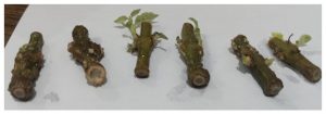
Among all treatments tested, T3 (BAP 3.0 + NAA 3.0 mg/L) produced the best overall organogenesis response, with maximum shoot proliferation, vigorous root induction, and formation of numerous axillary buds by day 13 shown in figure no 1.[2, 23] T4 (BAP 2.0 + NAA 3.0 mg/L) showed good shoot response but moderate rooting, indicating that elevated BAP levels synergize with NAA for balanced organogenesis.[21, 23] T1 at the lowest PGR combination produced the weakest response, confirming the need for adequate growth regulator concentrations for in vitro culture of this recalcitrant woody species.[21, 22].
Figure 2: Shoot and root multiplication visible in the photo (e.g., shoots only, roots,
callus, multiple shoots in culture bottles/test tubes).
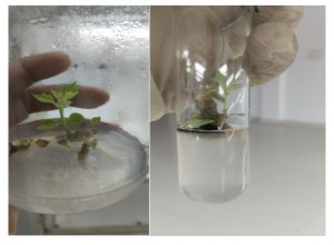
3.3 Hardening of Regenerated Plantlets: Rooted plantlets obtained from in vitro cultures were subjected to a stepwise acclimatization (hardening) protocol in a hardening chamber prior to ex vitro transfer. Plantlets were first transferred to plastic cups containing a sterile cocopeat:soil:sand (1:1:1) mixture and maintained under high humidity (80–90%) for one week, followed by gradual reduction in humidity over the subsequent two weeks. Survival rates of hardened plantlets were recorded. This process is critical to bridge the gap between heterotrophic in vitro conditions and autotrophic ex vitro growth shown in figure 2 (A & B).[2, 8, 22].
Figure 3: Hardening and acclimatization of tissue culture-raised plantlets of Gmelina arborea Roxb.: (A) plantlets transferred to cocopeat : soil : sand (1:1:1) mixture in root trainer under high humidity in hardening mist chamber; (B) successfully established plantlets under ex vitro conditions showing vigorous growth during lab-to-land transfer.

DISCUSSION
4.1 Surface Sterilization Efficacy: Contamination by fungi and bacteria is the most common cause of failure in plant tissue culture, particularly in woody species such as Gmelina arborea that harbour dense epiphytic and endophytic microbial communities.[8, 22] The present study evaluated a two-step sequential sterilization protocol combining Bavistin and HgCl₂, which proved highly effective for explant establishment.
Bavistin (Carbendazim) at 0.1% for 7 minutes provided optimal fungal control. This systemic fungicide inhibits β-tubulin polymerization in fungi, disrupting cell division and leading to fungal death. The inclusion of Tween-20 as a wetting agent enhanced the penetration and spread of the fungicide across the waxy surface of the explants.[8] These findings align with those of Anis et al. (2003)[1], who recommended Bavistin pre-treatment for surface sterilization of recalcitrant woody species prior to HgCl₂ application.
The subsequent application of 0.1% HgCl₂ for 2 minutes completely eliminated residual contamination. Mercury ions (Hg²⁺) are known to bind sulfhydryl groups in microbial proteins and enzymes, leading to irreversible denaturation and cell death. The 2-minute exposure was found to be sufficient for complete sterilization while preserving explant viability.[9] Prolonged exposure to HgCl₂ is known to cause oxidative stress, chlorophyll degradation, and impaired meristematic activity; thus, the optimized duration is of critical importance.[8, 22] Similar findings were reported by Husain et al. (2008)[9], who found that 0.1% HgCl₂ for 2 minutes produced the lowest contamination with satisfactory explant survival in Tectona grandis.
4.2 Effect of Plant Growth Regulators on Organogenesis: The organogenetic response of plant tissue cultures is critically governed by the type and concentration of plant growth regulators (PGRs) in the medium.[13, 23] In the present study, the combination of BAP (3 mg/L) + NAA (3 mg/L) on MS basal medium[14] produced the best shoot and root response in Gmelina arborea, with visible bud proliferation and root induction by day 13.
BAP (6-Benzylaminopurine), a synthetic adenine-type cytokinin, promotes axillary bud break, shoot proliferation, and cell division by activating cyclin-dependent kinases involved in the cell cycle. NAA (α-Naphthaleneacetic acid), a stable synthetic auxin, stimulates root initiation through polar transport and the activation of specific root-inducing genes.[13, 23] The synergistic interaction between BAP and NAA at equimolar concentrations (3:3 mg/L) created a hormonal balance that favoured both shoot and root development simultaneously — a response consistent with the concept of balanced organogenesis described by Skoog and Miller (1957)[23], who established the foundational auxin-to-cytokinin ratio model governing organogenesis in vitro.
These results confirm that the combined application of auxins and cytokinins significantly enhanced rooting and shooting in Gmelina arborea under in vitro conditions.[2, 22] Similar findings were reported by 21], who observed that balanced cytokinin–auxin combinations were essential for efficient micropropagation of recalcitrant hardwood species.
At sub-optimal concentrations (T1: BAP 2 + NAA 1 mg/L), growth regulator insufficiency likely limited cell division, resulting in poor organogenesis. At T4 (BAP 2 + NAA 3 mg/L), the auxin excess over cytokinin shifted the hormonal balance toward rooting at the expense of balanced shoot proliferation.[21, 23] This confirms the concentration-dependent and ratio-dependent nature of PGR-mediated organogenesis in Gmelina arborea.
4.3 Significance for Large-Scale Propagation: Gmelina arborea is a fast-growing, economically important tropical timber species with applications in agroforestry, pulpwood production, and social forestry programmes.[15, 16, 26] Conventional propagation through seeds suffers from low germination rates, genetic variability, and prolonged juvenile period.[12] The micropropagation protocol optimized in the present study employing a standardized sterilization regime followed by BAP + NAA-supplemented MS medium[14] provides a reliable, genotype-faithful, and season-independent method for clonal multiplication of elite trees.[22, 27] This assumes particular significance for the reforestation programmes of Madhya Pradesh, where high-quality planting material is in constant demand.[5]
CONCLUSION
The present investigation was undertaken to develop an efficient in vitro propagation protocol for Gmelina arborea Roxb. through nodal explants using Murashige and Skoog (1962) basal medium[14] supplemented with plant growth regulators. Based on the experimental results and discussion, the following conclusions are drawn:
- Sequential surface sterilization using 0.1% Bavistin for 7 minutes[1] followed by 0.1% HgCl₂ for 2 minutes[9] proved to be the most effective decontamination protocol, resulting in complete elimination of microbial contamination while maintaining explant viability.
- Among the PGR combinations evaluated, MS medium[14] supplemented with BAP 3.0 mg/L + NAA 3.0 mg/L (T3)[13, 23] elicited the highest organogenesis response, with maximum shoot proliferation, root induction, and formation of numerous axillary buds within 13 days of culture.
- The synergistic interaction between the cytokinin (BAP) and auxin (NAA) at equimolar concentrations (3:3 mg/L) provided the optimal hormonal balance for simultaneous shoot and root development, consistent with the Skoog–Miller model [23].
- Lower PGR concentrations (T1) were insufficient for adequate organogenetic response, while unequal ratios (T4) shifted the developmental pathway selectively toward shoots, indicating the importance of optimizing both concentration and ratio of PGRs.[21, 23]
- Hardening of regenerated plantlets in a controlled chamber facilitated successful acclimatization and transition to ex vitro conditions [8, 22], indicating the feasibility of the protocol for large-scale nursery application.
In conclusion, the optimized micropropagation protocol combining effective surface sterilization (Bavistin[1] + HgCl₂[ 9]) and an efficient PGR combination (BAP 3.0 + NAA 3.0 mg/L) [23] on MS basal medium[14] is highly suitable for reliable, rapid, and genetically uniform in vitro multiplication of Gmelina arborea. This protocol holds significant practical value for clonal forestry, conservation of plus trees, and large-scale production of quality planting material for afforestation programmes in India [16, 26].
ACKNOWLEDGEMENTS
The authors gratefully acknowledge the support provided by Shri Pradeep Vasudeva (IFS), PCCF & Director, State Forest Research Institute (SFRI), Jabalpur, Madhya Pradesh, India, for providing laboratory facilities and necessary support for this research work. The authors also express their sincere gratitude to Dr. Shailendra Singh Yadav, Supervisor, for his valuable technical guidance and assistance in the selection of plant materials throughout the course of this thesis investigation.
Conflict of Interest: The authors declare that there is no conflict of interest regarding the publication of this manuscript. The research was conducted in the absence of any commercial or financial relationships that could be construed as a potential conflict of interest.
REFERENCES
- Anis, M., Varshney, A., and Siddique, I. (2010). In vitro clonal propagation of Cassia angustifolia Agroforestry Systems, 78(2), 201–208. https://doi.org/10.1007/s10457-009-9239-8
- Arora, M. and Tamrakar, P. (2017). In vitro regeneration of Gmelina arboreathrough nodal explants: effect of plant growth regulators. Journal of Forestry Research, 28(3), 491–498. https://doi.org/10.1007/s11676-016-0316-y
- Beniwal, B.S. (1989). Silviculture of Tropical Trees. International Book Distributors, Dehradun. pp. 112–118.
- Bhatt, B.P., & Chauhan, D.S. (2003). Some multipurpose trees of the North-East India: cultural, medicinal and economic aspects. Journal of Bamboo and Rattan, 2(1), 1–12.
- Chaturvedi, A.N. (1993). Growth of Gmelina arborea in Uttar Pradesh. Van Vigyan, 31(1–4), 87–91.
- Chopra, R.N., Nayar, S.L., & Chopra, I.C. (1956). Glossary of Indian Medicinal Plants. Council of Scientific and Industrial Research, New Delhi. pp. 123–124.
- Gamble, J.S. (1902). A Manual of Indian Timbers. Sampson Low, Marston and Company, London. pp. 531–533.
- George, E.F., Hall, M.A., and De Klerk, G.J. (2008). Plant Propagation by Tissue Culture, Vol. 1: The Background (3rd ed.). Springer, Dordrecht. https://doi.org/10.1007/978-1-4020-5005-3
- Husain, M.K., Anis, M., and Shahzad, A. (2008). In vitro propagation of Indian Kino (PterocarpusmarsupiumRoxb.) using thidiazuron. In Vitro Cellular & Developmental Biology – Plant, 44(6), 522–528. https://doi.org/10.1007/s11627-008-9132-5
- Kadambi, K. (1954). On the ecology and silviculture of GmelinaarboreaRoxb.in the bamboo forests of Bhadravathi Division and notes on its distribution and cultivation in Mysore State. Indian Forester, 80(7), 358–374.
- Kirtikar, K.R., &Basu, B.D. (1935). Indian Medicinal Plants. Vol. III. Lalit Mohan Basu, Allahabad. pp. 1934–1937.
- Lauridsen, E.B., &Olesen, K. (1994). Experiences with Seed Sources of GmelinaarboreaRoxb.in Provenance Trials. Danish International Development Assistance (DANIDA), Forest Seed Centre, Denmark.
- Mitsukawa, N., Okumura, S., and Shirano, Y. (2001). Application of plant growth regulators in micropropagation. Plant Cell Reports, 20(4), 299–304.
- Murashige, T. and Skoog, F. (1962). A revised medium for rapid growth and bioassays with tobacco tissue cultures. Physiologia Plantarum, 15(3), 473–497. https://doi.org/10.1111/j.1399-3054.1962.tb08052.x
- National Research Council (NRC). (1983). Firewood Crops: Shrub and Tree Species for Energy Production. Vol. 2.National Academy Press, Washington D.C.
- Orwa, C., Mutua, A., Kindt, R., Jamnadass, R., & Anthony, S. (2009). Agroforestree Database: A Tree Reference and Selection Guide Version 4.0. World Agroforestry Centre, Nairobi, Kenya.
- Panshin, A.J., & de Zeeuw, C. (1980). Textbook of Wood Technology.4th ed. McGraw-Hill, New York.
- Patil, R., &Ghosh, S. (2012). Phytochemical analysis and in vitro antioxidant potential of Gmelina arboreabark extracts. Asian Journal of Plant Sciences, 11(4), 186–193.
- Pearson, R.S., & Brown, H.P. (1932). Commercial Timbers of India. Central Publication Branch, Government of India, Calcutta. pp. 617–621.
- Pullaiah, T. (2006). Encyclopaedia of World Medicinal Plants.Vol.2.Regency Publications, New Delhi. pp. 890–892.
- Rao, I.U. and Bhojwani, S.S. (1995). Tissue culture of forest trees. In: Bhojwani, S.S. (Ed.), Plant Tissue Culture: Applications and Limitations. Elsevier, Amsterdam, pp. 341–366.
- Rout, G.R., Samantaray, S., and Das, P. (2000). In vitro manipulation and propagation of medicinal plants. Biotechnology Advances, 18(2), 91–120. https://doi.org/10.1016/S0734-9750(99)00026-9
- Skoog, F. and Miller, C.O. (1957). Chemical regulation of growth and organ formation in plant tissues cultured in vitro. Symposia of the Society for Experimental Biology, 11, 118–130.
- Smitinand, T. (1980). Thai Plant Names.Royal Forest Department, Bangkok, Thailand.p. 174.
- Troup, R.S. (1921). The Silviculture of Indian Trees. Vol. II. Clarendon Press, Oxford. pp. 788–795.
- Verma, R.K., Verma, A., & Singh, R. (2015). Gmelina arborea: A review on its botany, agroforestry, wood properties, and medicinal applications. Journal of Forest Science, 61(8), 323–334.
- Zobel, B., Campinhos, E., &Ikemori, Y. (1983). Selecting and Breeding for Desirable Wood. Tappi Journal, 66(1), 70–74.