1Biotechnology Division, State Forest Research Institute, Jabalpur Madhya Pradesh, India
2Department of Microbiology, Govt. M.H. College of Home Science and Science for
Woman Autonomous, Jabalpur, Madhya Pradesh, India
Corresponding author email: biotech.yadav0@gmail.com
Article Publishing History
Received:
Accepted After Revision:
Santalum album Linn. (Indian Sandalwood; Family: Santalaceae) is a slow-growing, hemiparasitic tree species of exceptional economic, cultural, and pharmaceutical significance, classified as Vulnerable by the IUCN due to decades of over-exploitation. Its aromatic heartwood and essential oil rich in α-santalol and β-santalol accumulate only after 15–20 years of growth, making conventional seed-based and vegetative propagation methods inadequate for large-scale commercial and conservation demands. The present study was designed with two primary objectives: (i) to determine the optimal concentration and combination of Plant Growth Regulators (PGRs) for in vitro shoot proliferation, and (ii) to evaluate effective surface sterilization treatments to minimise microbial contamination during in vitro culture establishment of S. album nodal segment explants.
Nodal segment explants were collected from healthy, field-grown S. album trees at the State Forest Research Institute (SFRI), Jabalpur, Madhya Pradesh, India (January–March 2026). Explants were surface-sterilized using sequential treatments of 1% Bavistin (Carbendazim; 0–10 min) and Mercuric Chloride (HgCl₂; 0.0–0.5%; 1–5 min). Six combinations of 6-Benzylaminopurine (BAP: 0.0–4.0 mg/L) with Naphthalene Acetic Acid (NAA: 0.0–0.5 mg/L) were evaluated on Murashige and Skoog (MS, 1962) basal medium. Growth responses — initiation, proliferation, elongation, and final shoot status were recorded at 7, 14, 21, and 28 days each of culture. Hardened plantlets were acclimatized in a 1:1:1 (soil:vermicompost:sand) substrate under greenhouse conditions. Among all PGR treatments, the combination of 3.0 mg/L BAP with 0.5 mg/L NAA on MS basal medium yielded the maximum morphogenic response, recording 90–95% shoot initiation, 95–100% proliferation, and 90–95% elongation, with the production of healthy, vigorous plantlets. Supraoptimal BAP concentrations (4.0 mg/L) suppressed organised shoot development and induced undesirable callogenesis.
For surface sterilization, 1% Bavistin applied for 10 minutes reduced fungal contamination to 10% (from 100% in the untreated control), while 0.4% HgCl₂ for 2 minutes achieved the minimum contamination of 5–10% while maintaining full explant viability. Higher HgCl₂ concentrations (0.5%) caused phytotoxic tissue injury, paradoxically increasing contamination. The study establishes a reliable, reproducible micropropagation protocol for S. album using MS medium supplemented with 3.0 mg/L BAP + 0.5 mg/L NAA, with a sequential sterilization regimen of 1% Bavistin (10 min) followed by 0.4% HgCl₂ (2 min). This protocol offers a scalable foundation for clonal forestry, germplasm conservation, and commercial-scale production of Indian Sandalwood.
Santalum album Linn, Micropropagation, Nodal Explant, BAP, NAA, surface Sterilization, Mercuric Chloride, Carbendazim, Shoot Organogenesis, Plant Tissue Culture.
Yadav S. S, Rajak S, Bhagat P, Sandeep, Vasudeva P. In vitro Propagation of Santalum album Linn. (Indian Sandalwood): Optimization of Plant Growth Regulators and Surface Sterilization Protocols for Micropropagation. International Journal of Biomedical Research Science (IJBRS). 2026;02(2)
Yadav S. S, Rajak S, Bhagat P, Sandeep, Vasudeva P. In vitro Propagation of Santalum album Linn. (Indian Sandalwood): Optimization of Plant
Growth Regulators and Surface Sterilization Protocols for Micropropagation. International Journal of Biomedical Research Science (IJBRS). 2026; 02 (2). Available from: <a href=”https://shorturl.at/Oxgxo“>https://shorturl.at/Oxgxo</a>
INTRODUCTION
Santalum album Linn. (Indian sandalwood), belonging to the family Santalaceae, is one of the world’s most economically and culturally significant tree species. Widely acknowledged as the second most expensive wood globally, it has been prized for centuries for its intensely aromatic heartwood and essential oil, which find extensive applications in perfumery, cosmetics, pharmaceuticals, traditional medicine, and religious practices [10,4]. The species is native to the tropical dry deciduous forests of the Deccan Plateau in southern India, with Karnataka, Tamil Nadu, Andhra Pradesh, and Kerala together accounting for approximately 90% of its natural population [9]. It is also naturally distributed in Indonesia, Sri Lanka, and parts of the Pacific Islands.
The commercial value of S. album is largely attributed to its essential oil, which constitutes above 6% of the heartwood dry weight the highest among all Santalum species. The oil is predominantly composed of sesquiterpene alcohols, chiefly α-santalol and β-santalol, which together impart the characteristic woody-balsamic fragrance and are responsible for a range of pharmacological activities including antimicrobial, anti-inflammatory, antiviral, and chemopreventive properties [14,4,17]. Beyond its essential oil, phytochemical investigations of the heartwood, bark, leaves, fruits, seeds, and roots have revealed the presence of flavonoids, tannins, saponins, fatty acids (including the unique ximenynic acid), amino acids, and various terpenoids, underscoring the broad medicinal and industrial relevance of this species [13,5,16].
A distinguishing biological feature of S. album is its hemiparasitic nature. The tree possesses specialized root structures called haustoria, which enable it to attach to and draw water and inorganic nutrients from host plant root systems, while simultaneously being capable of conducting photosynthesis independently [7]. Compounding the challenges of natural propagation, S. album is a slow-growing species; the aromatic heartwood and essential oil accumulate only after 15–20 years of growth, and seed germination is often erratic due to dormancy issues, low viability, and high seedling mortality. These biological constraints, combined with decades of over-exploitation driven by the high market demand for sandalwood oil and timber, have led to a severe depletion of natural populations. Consequently, the International Union for Conservation of Nature (IUCN) has classified S. album as a Vulnerable species.
Given this scenario, conventional vegetative propagation methods such as grafting, budding, and layering have proven inadequate for meeting the large-scale demand for genetically uniform, high-quality planting material. Micropropagation through plant tissue culture offers a scientifically robust alternative, enabling the rapid mass multiplication of elite genotypes within a short time span while maintaining genetic fidelity [2]. Tissue culture techniques also facilitate the production of disease-free planting stock, conservation of threatened germplasm, and genetic improvement of economically important traits such as oil yield and growth rate. In vitro propagation of sandalwood was first attempted as early as [11] using mature endosperm on White’s medium, though callus proliferation was inconsistent. Since then, limited yet significant progress has been made in establishing standard micropropagation protocols for this species.
Two of the most critical determinants of successful in vitro propagation are the optimization of Plant Growth Regulators (PGRs) and the adoption of effective surface sterilization protocols. PGRs, including cytokinins (such as BAP and kinetin) and auxins (such as IBA and NAA), govern key developmental processes including axillary bud proliferation, shoot elongation, and adventitious root induction; however, their optimal concentration and combination are highly species- and explant-specific [6,3]. Concurrently, microbial contamination remains one of the foremost obstacles in tissue culture of woody species, particularly those like S. album that harbor endophytic microorganisms, making the selection of appropriate sterilization agents and treatment durations indispensable for culture establishment [1].
Despite the recognized importance of S. album and the urgent need for scalable propagation systems, standardized in vitro protocols for shoot proliferation and rooting remain insufficiently characterized. The present study, therefore, was designed with the dual objectives of: (i) determining the optimal concentration and combination of PGRs for shoot proliferation and root formation, and (ii) evaluating suitable sterilization treatments to minimize contamination during in vitro culture establishment of Santalum album Linn. The findings of this study are expected to contribute significantly toward developing a reliable micropropagation protocol that can support sustainable clonal forestry, conservation efforts, and commercial-scale production of Indian sandalwood.
Santalum album Linn. (Indian sandalwood), belonging to the family Santalaceae, is one of the world’s most economically and culturally significant tree species. Widely acknowledged as the second most expensive wood globally, it has been prized for centuries for its intensely aromatic heartwood and essential oil, which find extensive applications in perfumery, cosmetics, pharmaceuticals, traditional medicine, and religious practices [10,4]. The species is native to the tropical dry deciduous forests of the Deccan Plateau in southern India, with Karnataka, Tamil Nadu, Andhra Pradesh, and Kerala together accounting for approximately 90% of its natural population [9]. It is also naturally distributed in Indonesia, Sri Lanka, and parts of the Pacific Islands.
The commercial value of S. album is largely attributed to its essential oil, which constitutes above 6% of the heartwood dry weight the highest among all Santalum species. The oil is predominantly composed of sesquiterpene alcohols, chiefly α-santalol and β-santalol, which together impart the characteristic woody-balsamic fragrance and are responsible for a range of pharmacological activities including antimicrobial, anti-inflammatory, antiviral, and chemopreventive properties [14,4,17]. Beyond its essential oil, phytochemical investigations of the heartwood, bark, leaves, fruits, seeds, and roots have revealed the presence of flavonoids, tannins, saponins, fatty acids (including the unique ximenynic acid), amino acids, and various terpenoids, underscoring the broad medicinal and industrial relevance of this species [13,5,16].
A distinguishing biological feature of S. album is its hemiparasitic nature. The tree possesses specialized root structures called haustoria, which enable it to attach to and draw water and inorganic nutrients from host plant root systems, while simultaneously being capable of conducting photosynthesis independently [7]. Compounding the challenges of natural propagation, S. album is a slow-growing species; the aromatic heartwood and essential oil accumulate only after 15–20 years of growth, and seed germination is often erratic due to dormancy issues, low viability, and high seedling mortality. These biological constraints, combined with decades of over-exploitation driven by the high market demand for sandalwood oil and timber, have led to a severe depletion of natural populations. Consequently, the International Union for Conservation of Nature (IUCN) has classified S. album as a vulnerable species.
Given this scenario, conventional vegetative propagation methods such as grafting, budding, and layering have proven inadequate for meeting the large-scale demand for genetically uniform, high-quality planting material. Micropropagation through plant tissue culture offers a scientifically robust alternative, enabling the rapid mass multiplication of elite genotypes within a short time span while maintaining genetic fidelity [2]. Tissue culture techniques also facilitate the production of disease-free planting stock, conservation of threatened germplasm, and genetic improvement of economically important traits such as oil yield and growth rate. In vitro propagation of sandalwood was first attempted as early as [11]. using mature endosperm on White’s medium, though callus proliferation was inconsistent. Since then, limited yet significant progress has been made in establishing standard micropropagation protocols for this species.
Two of the most critical determinants of successful in vitro propagation are the optimization of Plant Growth Regulators (PGRs) and the adoption of effective surface sterilization protocols. PGRs, including cytokinins (such as BAP and kinetin) and auxins (such as IBA and NAA), govern key developmental processes including axillary bud proliferation, shoot elongation, and adventitious root induction; however, their optimal concentration and combination are highly species- and explant-specific [6,3]. Concurrently, microbial contamination remains one of the foremost obstacles in tissue culture of woody species, particularly those like S. album that harbor endophytic microorganisms, making the selection of appropriate sterilization agents and treatment durations indispensable for culture establishment [1].
Despite the recognized importance of S. album and the urgent need for scalable propagation systems, standardized in vitro protocols for shoot proliferation and rooting remain insufficiently characterized. The present study, therefore, was designed with the dual objectives of: (i) determining the optimal concentration and combination of PGRs for shoot proliferation and root formation, and (ii) evaluating suitable sterilization treatments to minimize contamination during in vitro culture establishment of Santalum album Linn. The findings of this study are expected to contribute significantly toward developing a reliable micropropagation protocol that can support sustainable clonal forestry, conservation efforts, and commercial-scale production of Indian sandalwood.
MATERIALS AND METHODS
2.1 Study Site and Experimental Period: The present investigation was carried out in the Tissue Culture, Biotechnology Division, State Forest Research Institute (SFRI), Jabalpur, Madhya Pradesh, India, during the period from January to March 2026. All in vitro experiments were performed under controlled aseptic conditions in the laboratory. The culture room was maintained at a temperature of 25 ± 2°C with a 16/8 h light/dark photoperiod provided by cool-white fluorescent lamps (2000–3000 lux) and a relative humidity of 50–70%.
2.2 Plant Material and Explant Selection: Healthy, disease-free nodal segments were collected as explants from mature Santalum album Linn. trees growing under natural field conditions. Selection of mother plants was made critically, considering the species’ recalcitrant nature and susceptibility to endophytic contamination. Young shoot tips and nodal segments (1.5–2.5 cm in length) were harvested from vigorous, actively growing branches during the months of April to June, when physiological activity is at its peak. Collection was carried out early in the morning to minimize wilting and physiological stress. Explants were immediately transported to the laboratory in sterile polythene bags containing moist sterile cotton, maintained in an ice box at approximately 4–10°C to preserve tissue viability.
2.3 Sterilization of Glassware and Equipment: All glassware including culture bottles, test tubes, conical flasks, volumetric flasks, Petri dishes, and measuring cylinders were thoroughly cleaned and processed using a two-step sterilization protocol. Initially, all items were placed in autoclavable disposal bags and subjected to steam sterilization (autoclaving) at 121°C and 15 psi pressure for 15–20 minutes. Following autoclaving, the glassware was immersed in 70% ethanol (v/v) for 10 minutes and subsequently rinsed thoroughly with double-distilled water (DDW). A second round of autoclaving was performed under the same conditions to ensure complete elimination of residual contamination. Prior to use, all sterilized glassware was placed inside the laminar airflow cabinet with UV light activated for 30 minutes to provide an additional layer of surface decontamination.
The following equipment was employed in the experimental procedures:
- Laminar airflow chamber (horizontal flow)
- Vertical autoclave (121°C, 15 psi)
- Hot air oven (180°C) for dry heat sterilization of heat-stable, moisture-sensitive items
- pH meter (calibrated with standard buffers at pH 4.0 and 7.0)
- Analytical balance and magnetic stirrer
- Microwave oven (for agar melting)
- Growth chamber with programmable photoperiod and temperature control
- Refrigerator (4°C) for hormone and stock solution storage
- Sterile culture bottles, test tubes, test tube stands, Petri plates, conical flasks, volumetric flasks, and filter paper
- Surgical blades, scalpels, forceps, and inoculation needles
- Non-absorbent cotton plugs, Parafilm, muslin cloth, and autoclave bags
2.4 Preparation of MS Basal Medium and Stock Solutions: The Murashige and Skoog (1962) basal medium was used throughout the study as the standard nutrient medium for in vitro culture of Santalum album. The MS medium was prepared from four concentrated stock solutions (Stocks A, B, C, and D) as described below. All chemicals used were of analytical reagent (AR) grade.
2.4.1 Preparation of Stock Solutions: Concentrated stock solutions were prepared separately for macronutrients, micronutrients, iron source, and vitamins, and stored at 4°C until use. The composition of each stock solution is detailed in Table 1.
Table 1. Chemical composition of Murashige and Skoog (1962) basal medium (per litre of final medium). *Note: For Santalum album, 50–100 mg/L Ascorbic Acid or 0.5–1.0 g/L Activated Charcoal may be added to combat phenolic oxidation.
| Stock | Component | Chemical Formula | Amount (mg/L) |
| Stock A (Macronutrients) | Ammonium Nitrate | NH₄NO₃ | 1650 |
| Potassium Nitrate | KNO₃ | 1900 | |
| Calcium Chloride | CaCl₂·2H₂O | 440 | |
| Stock B (Macronutrients) | Magnesium Sulphate | MgSO₄·7H₂O | 370 |
| Potassium Phosphate | KH₂PO₄ | 170 | |
| Stock C (Micronutrients) | Boric Acid | H₃BO₃ | 6.2 |
| Manganese Sulphate | MnSO₄·H₂O | 22.3 | |
| Zinc Sulphate | ZnSO₄·7H₂O | 8.6 | |
| Potassium Iodide | KI | 0.83 | |
| Sodium Molybdate | Na₂MoO₄·2H₂O | 0.25 | |
| Copper Sulphate | CuSO₄·5H₂O | 0.025 | |
| Cobalt Chloride | CoCl₂·6H₂O | 0.025 | |
| Stock D (Iron) | Ferrous Sulphate | FeSO₄·7H₂O | 27.8 |
| Disodium EDTA | Na₂EDTA·2H₂O | 37.3 | |
| Stock E (Vitamins) | Myo-inositol | — | 100.0 |
| Glycine | — | 2.0 | |
| Nicotinic Acid | — | 0.5 | |
| Pyridoxine HCl | — | 0.5 | |
| Thiamine HCl | — | 0.1 | |
| — | Sucrose (carbon source) | C₁₂H₂₂O₁₁ | 30,000 (3%) |
| — | Agar-Agar (gelling agent) | — | 8,000 (0.8%) |
| — | pH | — | 5.7–5.8 |
2.5 Preparation of Plant Growth Regulators (PGRs): Two plant growth regulators were used in the present study: 6-Benzylaminopurine (BAP), a cytokinin, and Naphthalene Acetic Acid (NAA), a synthetic auxin. Stock solutions of each PGR were prepared at a concentration of 1 mg/mL (1000 ppm). BAP was dissolved in a few drops of 0.1 N NaOH and then made up to volume with DDW. NAA was dissolved in a minimum quantity of 95% ethanol and then diluted with DDW to the required volume. All hormone stock solutions were filter-sterilized (0.22 µm Millipore filter) and stored in amber-coloured glass vials at 4°C in a refrigerator to prevent photodegradation and degradation. Appropriate aliquots were added to the autoclaved and cooled medium at 45–50°C just before pouring.
2.6 Surface Sterilization of Explants: Surface sterilization of nodal segment explants was carried out following a sequential protocol designed to eliminate surface-borne and endophytic contaminants while minimizing damage to the explant tissue. The protocol was as follows:
Step 1 — Pre-washing: Explants were gently washed under running tap water for 20–30 minutes to remove dust, debris, and loosely adhering microorganisms.
Step 2 — Tween-20 Treatment: The explants were immersed in a 10% Tween-20 (polyoxyethylene sorbitan monolaurate) solution for 10 minutes with continuous gentle agitation to remove surface-adhering contaminants and reduce surface tension, followed by thorough rinsing with distilled water 4–5 times.
Step 3 — Fungicide Treatment: Explants were then treated with 1% Bavistin (Carbendazim, a systemic fungicide) solution for 30 minutes with occasional shaking to prevent fungal contamination. This was followed by 4–5 washes with sterile distilled water.
Step 4 — Surface Sterilant Treatment (HgCl₂ / NaOCl): All subsequent steps were performed inside the laminar airflow cabinet under aseptic conditions. The explants were treated with either Mercuric Chloride (HgCl₂) at concentrations of 0.05% and 0.1% (w/v), or Sodium Hypochlorite (NaOCl) at concentrations of 0.25% and 4% (v/v), for varying time intervals of 5, 7, 10, and 15 minutes. This constituted a factorial treatment design to determine the most effective sterilization regimen that minimizes contamination while maintaining maximum explant viability.
Step 5 — Rinsing: Following sterilant treatment, explants were rinsed thoroughly 3–4 times with sterile double-distilled water to completely eliminate residual sterilizing agents, which could otherwise be phytotoxic.
2.7 Inoculation of Explants: Inoculation of explants was performed aseptically inside a horizontal laminar airflow chamber. Prior to inoculation, the laminar airflow cabinet was exposed to UV irradiation for 30 minutes. The working surface was then swabbed thoroughly with 70% ethanol (v/v). The researcher wore sterile gloves and face mask throughout the procedure. All instruments including forceps, scalpels, and needles were flamed using a spirit lamp and allowed to cool before use. Culture vessels containing the prepared MS medium were briefly flamed at their mouths before opening.
Sterilized explants were trimmed to a uniform length of 1.5–2.0 cm using a sterile scalpel, ensuring that each segment contained at least one node. The explants were then inoculated vertically into the culture medium, with the basal end embedded in the agar. The mouths of the culture vessels were re-flamed before capping. All culture vessels were sealed with Parafilm or autoclavable caps to maintain sterility and labelled with date of inoculation, treatment details, and medium composition.
2.8 Incubation Conditions: After inoculation, all culture vessels were transferred to the culture room and maintained under the following controlled environmental conditions throughout the experimental period:
- Temperature: 25 ± 2°C
- Photoperiod: 16 hours light / 8 hours dark
- Light intensity: 2000–3000 lux (cool-white fluorescent lamps)
- Relative humidity: 50–70%
Cultures were inspected regularly at intervals of 48–72 hours to record contamination, browning, and morphogenic responses including shoot initiation, shoot elongation, callus formation, and root induction.
2.9 Hardening and Ex Vitro Acclimatization: Well-rooted plantlets showing adequate shoot development were selected for ex vitro establishment. Prior to transplanting, plantlets were rinsed thoroughly with sterile distilled water to remove residual medium and agar from the root system, which can otherwise promote microbial growth in soil. Plantlets were then transferred to paper cups containing a sterile potting mixture of soil, vermicompost, and sand in the ratio of 1:1:1 (v/v/v). Transplanted plantlets were maintained initially in the greenhouse under 60–70% relative humidity and light shade to reduce transplant shock.
For the first 15 days, plantlets were fertigated with half-strength MS basal liquid medium to support nutritional needs during the transition from in vitro to ex vitro conditions. After successful acclimatization, hardened plantlets were transferred to 6 cm diameter pots and subsequently to larger 14 cm diameter pots containing the same substrate mixture. The survival percentage of acclimatized plantlets was recorded at 15 and 30 days after transplanting.
2.10 Experimental Design and Statistical Analysis: The experiments were laid out in a Completely Randomized Design (CRD). Each treatment was replicated three times (n = 3), with five culture tubes per replication. Observations were recorded for: (i) percentage of contamination-free cultures, (ii) percentage of explant survival, (iii) days to shoot initiation, (iv) number of shoots per explant, (v) shoot length (cm), and (vi) percentage of rooting. Data were expressed as mean ± standard error (SE) and subjected to one-way analysis of variance (ANOVA). Means were compared using Duncan’s Multiple Range Test (DMRT) at p ≤ 0.05.
RESULTS AND DISCUSSION
3.1 Effect of BAP and NAA on In Vitro Shoot Growth of Santalum album Linn: The effect of varying concentrations of 6-Benzylaminopurine (BAP) in combination with Naphthalene Acetic Acid (NAA) on the in vitro growth response of Santalum album nodal segment explants was evaluated at four progressive time intervals: 14 days (initiation), 28 days (proliferation), 42 days (elongation), and 60 days (final shoot status). The data are presented in Table 2. The results clearly demonstrated that the growth response was significantly dependent on the concentration and combination of PGRs supplemented to the MS basal medium.
3.1.1 Control Treatment (T0): Explants maintained on hormone-free MS basal medium exhibited a no morphogenic response throughout the culture period. Initiation percentage remained low (15–25%), proliferation was minimal (20–30%), and elongation was negligible (25–35%). No organised shoot development was recorded at 14 days. These findings confirm that the endogenous hormone levels in the excised nodal explants of Santalum album are insufficient to sustain active organogenesis under in vitro conditions, and exogenous PGR supplementation is indispensable for culture establishment.
3.1.2 Low Concentration Treatment (T1): The addition of low concentrations of BAP and NAA resulted in a moderate improvement in growth response. Initiation frequency increased to 40–50%, while proliferation and elongation reached 50–60% and 55–65%, respectively. However, the shoot formation observed at 28 days remained weak and poorly developed, indicating that the hormonal stimulus provided at this level was insufficient to induce robust organogenesis.
3.1.3 Moderate Concentration Treatments (T2): A significant improvement in growth response was observed as the BAP concentration was increased to 1.0 mg/L combined with 0.2 mg/L NAA (T2). Initiation frequency improved to 60–70%, proliferation to 70–80%, and elongation to 75–85%, with moderate shoot formation recorded. Further increase to 2.0 mg/L BAP with 0.3 mg/L NAA (T3) resulted in a substantial enhancement, with initiation reaching 80–90%, proliferation 85–95%, and elongation 85–90%. Good shoot multiplication was recorded under this treatment, demonstrating a positive dose-response relationship at intermediate PGR concentrations.
3.1.4 Optimal Treatment (T4) ★: The maximum and most desirable morphogenic response was obtained at 3.0 mg/L BAP combined with 0.5 mg/L NAA. This treatment recorded the highest initiation percentage (90–95%), proliferation rate (95–100%), and elongation (90–95%). Shoots produced were healthy, well-developed, green, and exhibited vigorous growth. This combination proved to be the most effective for in vitro shoot regeneration of Santalum album in the present study and is recommended as the optimal PGR regime for shoot induction.
3.1.5 High Concentration Treatment (T5): A further increase in BAP concentration to 4.0 mg/L, while maintaining NAA at 0.5 mg/L, resulted in a decline in organised shoot formation. Initiation and proliferation decreased to 50–60% and 55–65% respectively, and extensive callus formation was observed at the base of explants, with poor shoot development at 28 days. This inhibitory effect at supra-optimal cytokinin levels is a well-documented phenomenon in woody plant tissue culture, where excessively high BAP concentrations suppress organised shoot morphogenesis and redirect developmental pathways toward unorganised callus proliferation [3].
Table 2. Effect of BAP and NAA combinations on in vitro growth response of Santalum album Linn. at different time intervals. ★ Optimal treatment.
| Treatment | BAP (mg/L) | NAA (mg/L) | 14 Days Initiation (%) | 28 Days Proliferation (%) | 42 Days Elongation (%) | 60 Days — Final Status |
| T0 | 0.0 | 0.0 | 15–25 | 20–30 | 25–35 | Very poor; no shoot development |
| T1 | 0.5 | 0.1 | 40–50 | 50–60 | 55–65 | Weak shoot formation |
| T2 | 1.0 | 0.2 | 60–70 | 70–80 | 75–85 | Moderate shooting |
| T3 | 2.0 | 0.3 | 80–90 | 85–95 | 85–90 | Good shoot proliferation |
| T4 ★ | 3.0 | 0.5 | 90–95 | 95–100 | 90–95 | Excellent; vigorous healthy plantlets |
| T5 | 4.0 | 0.5 | 50–60 | 55–65 | 60–70 | Callus formation; poor organised shooting |
The results of the present study were in agreement with established principles of plant growth regulator interactions in tissue culture systems. Cytokinins such as BAP are well known to promote cell division and axillary bud activation, while auxins such as NAA facilitate cell elongation, differentiation, and root initiation [6]. In the present investigation, the combined application of BAP and NAA demonstrated a clear synergistic effect on shoot initiation, proliferation, and elongation, with the optimal response achieved at a BAP:NAA ratio of 6:1 (3.0 mg/L : 0.5 mg/L).
These findings were consistent with earlier reports on recalcitrant woody species, where a balanced cytokinin-to-auxin ratio is critical for achieving maximum shoot regeneration frequency. Deviations from this optimal balance either through insufficient hormone supply (as in T0 and T1) or through supraoptimal cytokinin levels (as in T5) resulted in markedly reduced regeneration efficiency or abnormal callogenesis. The dominance of BAP in this study for shoot induction corroborates reports by [12]. and subsequent workers on Santalum album, who identified BAP as the primary shoot-inducing cytokinin in this species.
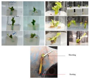
Figure 1: In vitro morphogenic response of Santalum album Linn. nodal explants cultured on MS medium supplemented with varying concentrations of BAP and NAA. (A) Initial bud break at day 14; Early shoot emergence with axillary bud activation; Shoot elongation with leaf primordia development; Progressive shoot proliferation and root initiation under auxin-dominant treatment conditions.
3.2 Effect of Bavistin (Carbendazim) Concentration and Exposure Duration on Explant Sterilization: Surface sterilization using the systemic broad-spectrum fungicide Bavistin (1% Carbendazim) was evaluated at six exposure durations (0, 2, 4, 6, 8, and 10 minutes) to determine the optimal treatment for controlling fungal contamination of Santalum album nodal segment explants. Results are presented in Table 3.
3.2.1 Control (T0 — 0 minutes): In the absence of Bavistin treatment, 100% contamination was recorded in all explants, predominantly by fungal microorganisms. This confirms the presence of a heavy endophytic and epiphytic microbial load on the surface of field-collected sandalwood explants, making pre-treatment with a fungicide an absolute necessity prior to surface sterilization.
3.2.2 Progressive Reduction in Contamination with Increasing Exposure Duration: A consistent and progressive decline in contamination percentage was observed with increasing Bavistin exposure duration. At 2 minutes (T1), contamination was reduced to 80%, indicating only partial removal of surface fungi. At 4 minutes (T2), the contamination declined further to 60%, and at 6 minutes (T3) to 40%, demonstrating that increased contact time markedly improves the fungicidal efficacy of Bavistin. A significant reduction was achieved at 8 minutes (T4), where contamination dropped to 20%, with the majority of explants surviving in viable condition.
3.2.3 Optimal Treatment (T5 — 1% Bavistin for 10 minutes) ★: The lowest contamination rate of 10% was recorded at 10 minutes of exposure to 1% Bavistin, representing the most effective duration tested. This treatment was selected as optimal as it provided the best balance between maximum contamination control and acceptable explant survival and regeneration potential. Prolonged Bavistin exposure beyond 10 minutes was not tested in the present study; however, it is well established that excessive fungicide treatment can induce chemical phytotoxicity, leading to tissue browning, reduced viability, and delayed culture establishment [1].
The progressive reduction in fungal contamination with increasing Bavistin exposure duration can be attributed to greater contact time between Carbendazim molecules and fungal cell wall components, enabling more complete inhibition of beta-tubulin polymerisation the primary mode of action of benzimidazole fungicides. The results demonstrate that 8–10 minutes of treatment with 1% Bavistin represents the optimal sterilization window for Santalum album, providing near-complete fungal decontamination while preserving explant integrity. These findings are consistent with reports from woody plant tissue culture where pre-treatment with systemic fungicides significantly reduces endophyte-mediated contamination that would otherwise evade conventional surface sterilants such as NaOCl or HgCl2.
Table 3. Effect of 1% Bavistin (Carbendazim) exposure duration on contamination percentage of Santalum album Linn. nodal segment explants. ★ Optimal treatment.
| Treatment | Bavistin Conc. (%) | Exposure Duration (min) | Contamination (%) | Observations |
| T0 | 1% | 0 (Control) | 100 | Complete fungal colonisation; no culture establishment possible |
| T1 | 1% | 2 | 80 | Minimal reduction; majority of explants heavily contaminated |
| T2 | 1% | 4 | 60 | Moderate reduction; partial fungal control observed |
| T3 | 1% | 6 | 40 | Improved sterilization; more than half of explants contamination-free |
| T4 | 1% | 8 | 20 | Significant decline in contamination; most explants viable |
| T5★ | 1% | 10 | 10 | Optimal: minimum contamination with acceptable explant survival |
3.3 Effect of HgCl2 Concentration and Exposure Duration on Explant Sterilization: The effect of varying concentrations of Mercuric Chloride (HgCl₂) combined with different exposure durations on the sterilization efficacy and explant survival of Santalum album nodal segments was evaluated. Five treatment concentrations (0.1–0.5%) with inversely decreasing exposure durations (5 to 1 min) were tested alongside an untreated control. Results are presented in Table 4.
3.3.1 Control (T0 — 0.0% HgCl2): As expected, the untreated control recorded 100% contamination, confirming the necessity of chemical sterilization for field-collected explants of Santalum album. Both bacterial and fungal contaminants were observed, rendering culture establishment impossible without appropriate sterilization.
3.3.2 Progressive Effect of Increasing HgCl₂ Concentration: Contamination progressively decreased as HgCl₂ concentration was increased from 0.1% to 0.4%. At 0.1% HgCl₂ for 5 minutes (T1), contamination was reduced to 60–70%, indicating only partial surface sterilization. Increasing concentration to 0.2% (T2, 4 min) further reduced contamination to 30–40%, and at 0.3% (T3, 3 min), contamination was markedly reduced to 10–20%, with most explants appearing healthy and viable.
3.3.3 Optimal Treatment (T4) ★: The most effective sterilization was achieved at 0.4% HgCl₂ for 2 minutes, which reduced contamination to a minimum of 5–10% the lowest recorded in this experiment. Explants treated under this condition remained viable, showed no visible signs of tissue browning or damage, and exhibited successful morphogenic response upon transfer to culture medium. This treatment is therefore recommended as the optimal HgCl₂ protocol for Santalum album nodal segment sterilization.
3.3.4 Effect of Higher Concentration (T5): At 0.5% HgCl₂, despite shorter exposure duration of only 1 minute, contamination marginally increased to 10–15% compared to T4. This paradoxical increase is attributable to HgCl₂-induced phytotoxicity at higher concentrations, which causes cellular damage and tissue injury in the explant, thereby compromising the natural antimicrobial barrier of the plant tissue and potentially facilitating secondary microbial invasion. This observation underscores the importance of selecting a moderate HgCl₂ concentration rather than maximising concentration in isolation.
HgCl₂ is a broad-spectrum biocide effective against bacteria, fungi, and spores due to its strong protein-denaturing and enzyme-inhibiting properties, acting primarily through binding to sulfhydryl groups of microbial proteins. The present study confirms that 0.3–0.4% HgCl₂ represents the optimal concentration range for Santalum album, providing effective sterilization while maintaining explant viability. The inverse relationship between concentration and exposure time (higher concentration used for shorter duration) is a standard approach that minimises tissue toxicity while maximising sterilization efficacy. These results align with reports by [8]. and others working on recalcitrant tropical tree species, where 0.1–0.5% HgCl₂ for 2–10 minutes has been found most effective.
Table 4. Effect of HgCl₂ concentration and exposure duration on contamination of Santalum album Linn. nodal segment explants. ★ Optimal treatment.
| Treatment | HgCl₂ Conc. (%) | Exposure Duration (min) | Contamination (%) | Observations |
| T0 | (Control)0.0 | 0 | 100 | No sterilization; complete microbial colonisation |
| T1 | 0.1 | 5 | 60–70 | Partial removal of surface microorganisms |
| T2 | 0.2 | 4 | 30–40 | Improved sterilization; moderate contamination remaining |
| T3 | 0.3 | 3 | 10–20 | Good sterilization; low contamination; explants healthy |
| T4 ★ | 0.4 | 2 | 5–10 | Optimal: maximum sterilization; explants viable and healthy |
| T5 | 0.5 | 1 | 10–15 | Marginal rise in contamination; tissue injury at this conc. |
CONCLUSION
The present investigations were on the in vitro propagation of Santalum album Linn. (Indian Sandalwood) through nodal segment culture on MS basal medium demonstrated that micropropagation was a highly effective and scalable approach for the multiplication and conservation of this economically critical and IUCN-vulnerable tree species. The study successfully addressed the two core objectives: optimisation of plant growth regulator concentrations for shoot proliferation and root formation, and evaluation of effective surface sterilization protocols to minimise contamination.
With respect to the first objective, the investigation conclusively established that the in vitro growth and morphogenesis of Santalum album are significantly regulated by the type, concentration, and balance of exogenous plant growth regulators. Among all tested combinations, 3.0 mg/L BAP supplemented with 0.5 mg/L NAA on MS basal medium yielded the maximum shoot response recording 90–95% initiation frequency, 95–100% proliferation, and 90–95% elongation at 28 days with the production of healthy, vigorous, well-developed plantlets. This combination is recommended as the optimal PGR regime for routine in vitro shoot regeneration of Santalum album. Supraoptimal BAP concentrations (4.0 mg/L) were found to promote callogenesis at the expense of organised shoot formation, confirming that a balanced cytokinin-to-auxin ratio is critical for directed organogenesis.
With respect to the second objective, two sequential sterilization protocols were evaluated. For fungal decontamination, 1% Bavistin (Carbendazim) applied for 10 minutes proved optimal, reducing fungal contamination from 100% (untreated control) to 10% while maintaining explant viability. For broad-spectrum sterilization of bacterial and fungal contaminants, 0.4% Mercuric Chloride (HgCl₂) applied for 2 minutes was found most effective, reducing contamination to 5–10% the minimum recorded in this study without causing significant tissue toxicity. Higher HgCl₂ concentrations (0.5%) resulted in a paradoxical marginal increase in contamination attributable to phytotoxic tissue injury, highlighting the critical importance of concentration optimization rather than maximisation.
The [6] basal medium, supplemented with sucrose as carbon source, proved to be the most suitable nutritional base for Santalum album in vitro culture. The addition of ascorbic acid or activated charcoal to the medium was found beneficial in counteracting the characteristic phenolic browning associated with this high-tannin species. Successful hardening and acclimatization of rooted plantlets was achieved using a 1:1:1 (soil:vermicompost:sand) substrate under greenhouse conditions, with half-strength MS liquid medium fertigation supporting the transition from in vitro to ex vitro conditions.
Taken together, the findings of this study provide a standardized, reproducible protocol for the micropropagation of Santalum album Linn. that can serve as a reliable foundation for commercial-scale clonal propagation, conservation of superior genotypes, and germplasm banking programs. The protocol developed is particularly significant given the increasing demand for sandalwood and the limited natural regeneration capacity of this slow-growing hemiparasitic species. Future research should focus on optimisation of rooting induction using IBA, genetic fidelity assessment of micropropagated plantlets through molecular markers, and evaluation of oil content in tissue culture-raised trees relative to naturally grown specimens.
ACKNOWLEDGEMENTS
The authors gratefully acknowledge the support provided by Shri Pradeep Vasudeva (IFS) PCCF & Director, State Forest Research Institute (SFRI), Jabalpur, Madhya Pradesh, India, and author also thankful to Dr. Shailendra Singh Yadav for providing plant material, laboratory facilities, and technical assistance throughout the course of this investigation.
Conflict of Interest: The authors declare that there is no conflict of interest regarding the publication of this manuscript. The research was conducted in the absence of any commercial or financial relationships that could be construed as a potential conflict of interest.
REFERENCES
- Bhojwani, S.S. and Razdan, M.K. (1996). Plant Tissue Culture: Theory and Practice. Elsevier Science, Amsterdam.
- Chalak, L. and Rogers, S.M.D. (1990). Micropropagation of sandalwood. Scientia Horticulturae, 43(3-4): 337–343.
- George, E.F., Hall, M.A. and De Klerk, G.J. (2008). Plant Propagation by Tissue Culture, 3rd edition. Springer, Netherlands.
- Howes, M.R., Simmonds, M.S.J. and Kite, G.C. (2004). Evaluation of the quality of sandalwood essential oils by gas chromatography–mass spectrometry. Journal of Chromatography A, 1028(2): 307–312.
- Liu, H.J., Fan, R.H. and Mao, Z.H. (1996). Fatty acid composition of Santalum album seed oil. Journal of the American Oil Chemists’ Society, 73(9): 1193–1195.
- Murashige, T. and Skoog, F. (1962). A revised medium for rapid growth and bioassays with tobacco tissue cultures. Physiologia Plantarum, 15(3): 473–497.
- Nagaveni, H.C. and Srimathi, R.A. (1985). Studies on the morphological and anatomical characters of Santalum album Linn. Indian Forester, 111: 743–749.
- Patel, R. and Rao, S. (2013). Surface sterilization protocols for in vitro propagation of Santalum album. Indian Journal of Biotechnology, 12(2): 245–250.
- Radomiljac, A.M., McComb, J.A. and Pate, J.S. (1998). Gas exchange and water relations of the root hemi-parasite Santalum album L. Annals of Botany, 82: 207–215.
- Rai, S.N. (1990). Status and cultivation of sandalwood in India. In: Proceedings of the Symposium on Sandalwood in the Pacific. USDA Forest Service, Pacific Southwest Research Station, Honolulu, pp. 66–71.
- Rangaswamy, N.S. and Rao, P.S. (1963). Experimental studies on Santalum album: Cultivation of endosperm in vitro. Phytomorphology, 13: 450–454.
- Rao, P.S., Bapat, V.A. and Mhatre, M. (2005). Santalum album: Biotechnological approaches for propagation and improvement. In: Biotechnology in Agriculture and Forestry, vol. 56. Springer, Berlin, pp. 285–302.
- Shankaranarayana, K.H., Ayyar, M.S. and Theagarajan, K.S. (1980a). Tannins of Santalum album heartwood. Indian Forester, 106(5): 355–358.
- Verghese, J., Joy, M.T. and Retnam, M. (1990). East Indian sandalwood oil — A review. Flavor and Fragrance Journal, 5(3): 107–122.
- Yan, H., Chen, M., Zhao, Z. and Hu, Z. (2013). Further studies on flavonoid constituents of Santalum album heartwood. Asian Pacific Journal of Tropical Biomedicine, 3(4): 307–310.
- Yan, H., Zhong, G., Zhao, Z. and Hu, Z. (2011). Flavonoids from Santalum album heartwood. Natural Product Research, 25(16): 1527–1532.
- Zhang, X., Niu, S. and Liu, Z. (2012). Chemical composition and antioxidant properties of Santalum album essential oil. Journal of Medicinal Plants Research, 6(14): 2935–2941.